Copyright
©The Author(s) 2021.
World J Gastroenterol. Jul 21, 2021; 27(27): 4322-4341
Published online Jul 21, 2021. doi: 10.3748/wjg.v27.i27.4322
Published online Jul 21, 2021. doi: 10.3748/wjg.v27.i27.4322
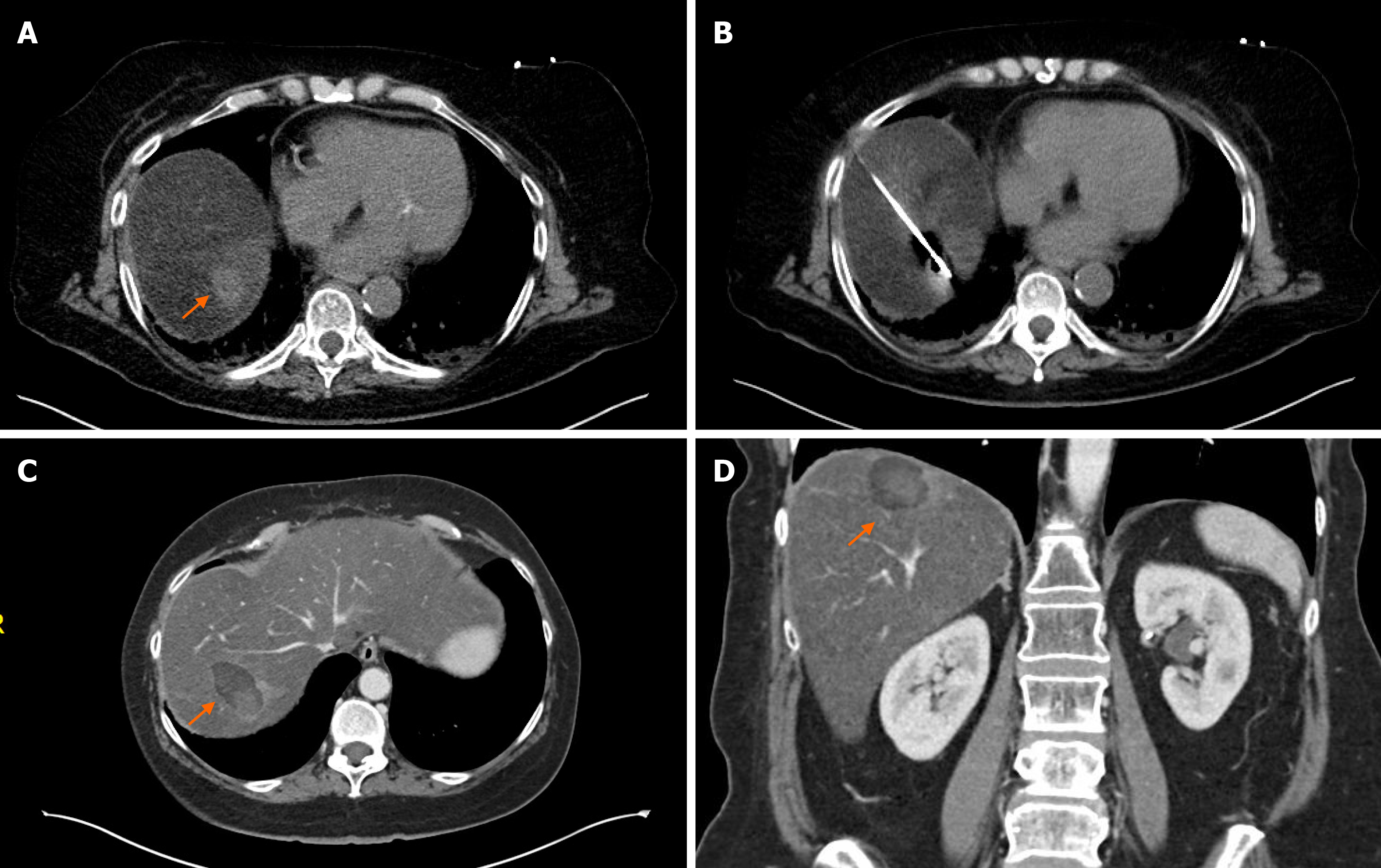
Figure 3 Percutaneous computed tomography-guided microwave ablation of a solitary pancreatic adenocarcinoma liver metastasis.
A: 62-year-old female presented with a 2 cm solitary liver metastasis (orange arrow), 9 mo after the surgical resection of the primary tumor from the pancreatic head; B: Intraprocedural image shows the microwave antenna in the liver metastasis; C and D: Axial and coronal contrast-enhanced computed tomography images 1 mo after the ablation demonstrates a 4.3 Í 2.7 cm hypodense ablation zone (orange arrows). There was no evidence of residual or new metastases in the liver. Patient is currently on systemic therapy 2 mo after the ablation without evidence of disease.
- Citation: Bibok A, Kim DW, Malafa M, Kis B. Minimally invasive image-guided therapy of primary and metastatic pancreatic cancer. World J Gastroenterol 2021; 27(27): 4322-4341
- URL: https://www.wjgnet.com/1007-9327/full/v27/i27/4322.htm
- DOI: https://dx.doi.org/10.3748/wjg.v27.i27.4322


