Copyright
©The Author(s) 2021.
World J Gastroenterol. Jul 14, 2021; 27(26): 4172-4181
Published online Jul 14, 2021. doi: 10.3748/wjg.v27.i26.4172
Published online Jul 14, 2021. doi: 10.3748/wjg.v27.i26.4172
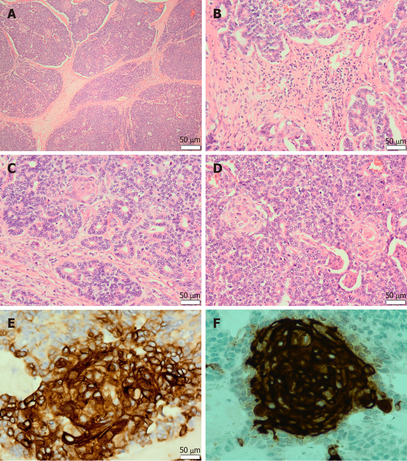
Figure 1 Pancreatoblastoma.
A: The tumour is composed of lobules separated by dense fibrous bands, imparting a geographic low power appearance [Haematoxylin and Eosin (H&E) staining, 40 ×]; B: The dense fibrous bands between the lobules are composed of spindled cells with varying amounts of collagen (H&E staining, 200 ×); C: The tumour predominantly shows acinar differentiation. The acinar units are composed of neoplastic cells arranged around central lumina (H&E staining, 200 ×); D: The tumour shows characteristic squamoid nests. Squamoid nests are large islands of plump epithelioid cells with abundant eosinophilic cytoplasm (H&E staining, 200 ×); E: The squamoid nests are immunoreactive for AE1/AE3 (400 ×); F: The tumour shows immunolabeling for CD10 limited to the squamoid nests (400 ×).
- Citation: Omiyale AO. Adult pancreatoblastoma: Current concepts in pathology. World J Gastroenterol 2021; 27(26): 4172-4181
- URL: https://www.wjgnet.com/1007-9327/full/v27/i26/4172.htm
- DOI: https://dx.doi.org/10.3748/wjg.v27.i26.4172


