Copyright
©The Author(s) 2021.
World J Gastroenterol. Jul 14, 2021; 27(26): 4143-4159
Published online Jul 14, 2021. doi: 10.3748/wjg.v27.i26.4143
Published online Jul 14, 2021. doi: 10.3748/wjg.v27.i26.4143
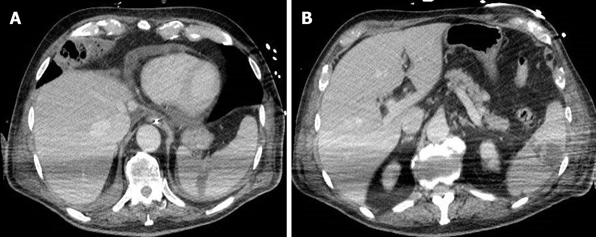
Figure 13 A 77-year-old man with abdominal tenderness.
A: Contrast-enhanced portal-venous phase computed tomography image of the abdomen demonstrated a wedge-shaped low-attenuation area at the level of the spleen, typical of infarction. Pericardial effusion was also present; B: A further rounded low-attenuation area with peripheral localization was present in a lower portion of the spleen on contrast-enhanced portal-venous phase computed tomography scan.
- Citation: Boraschi P, Giugliano L, Mercogliano G, Donati F, Romano S, Neri E. Abdominal and gastrointestinal manifestations in COVID-19 patients: Is imaging useful? World J Gastroenterol 2021; 27(26): 4143-4159
- URL: https://www.wjgnet.com/1007-9327/full/v27/i26/4143.htm
- DOI: https://dx.doi.org/10.3748/wjg.v27.i26.4143


