Copyright
©The Author(s) 2021.
World J Gastroenterol. Jul 14, 2021; 27(26): 4143-4159
Published online Jul 14, 2021. doi: 10.3748/wjg.v27.i26.4143
Published online Jul 14, 2021. doi: 10.3748/wjg.v27.i26.4143
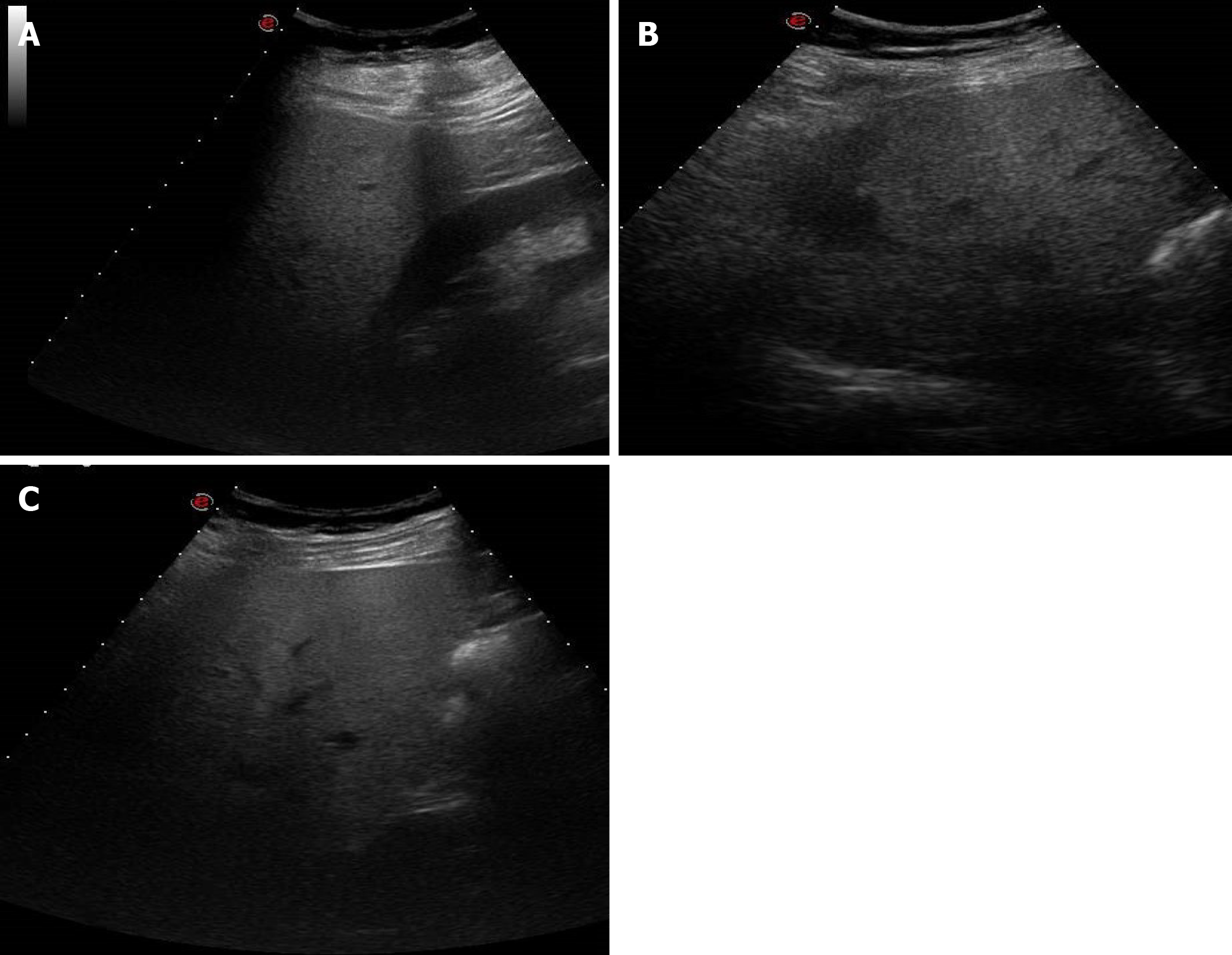
Figure 8 A 38-year-old man with abdominal discomfort.
A: Abdominal ultrasound image demonstrated hepatic steatosis as an increase in echogenicity compared to the renal cortex; B: Abdominal ultrasound image demonstrated hepatic steatosis as loss of physiological hyperechogenicity of the wall of the portal branches; C: Abdominal ultrasound image demonstrated hepatic steatosis as posterior attenuation of the ultrasonic beam with failure to visualize the diaphragm.
- Citation: Boraschi P, Giugliano L, Mercogliano G, Donati F, Romano S, Neri E. Abdominal and gastrointestinal manifestations in COVID-19 patients: Is imaging useful? World J Gastroenterol 2021; 27(26): 4143-4159
- URL: https://www.wjgnet.com/1007-9327/full/v27/i26/4143.htm
- DOI: https://dx.doi.org/10.3748/wjg.v27.i26.4143


