Copyright
©The Author(s) 2021.
World J Gastroenterol. Jul 7, 2021; 27(25): 3888-3900
Published online Jul 7, 2021. doi: 10.3748/wjg.v27.i25.3888
Published online Jul 7, 2021. doi: 10.3748/wjg.v27.i25.3888
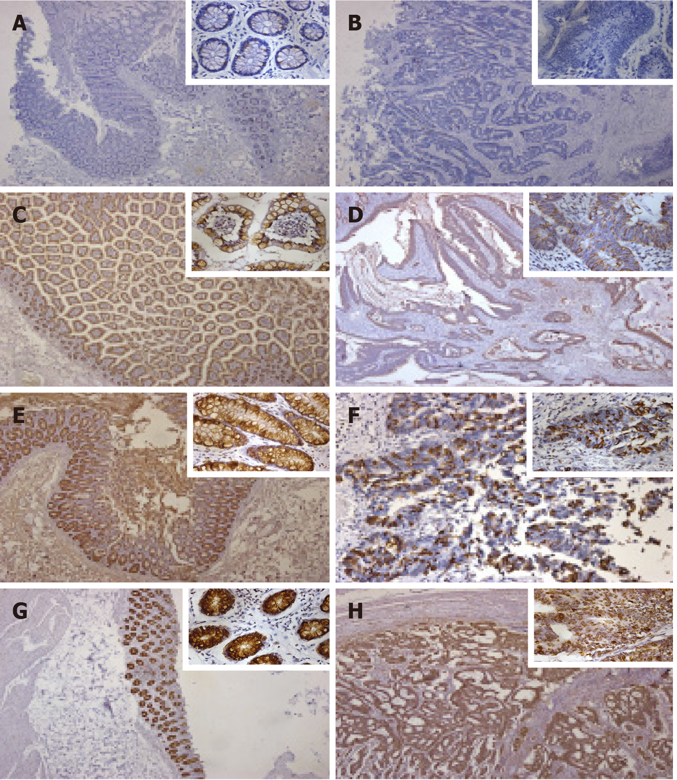
Figure 2 Representative pictures of immunohistochemical staining for mucin 2 in normal and cancer tissues of patients with colorectal cancer.
A and B: Negative expression; C and D: Weak expression; E and F: Moderate expression; G and H: Strong expression. A, C, E, and G: Normal tissue; B, D, F, and H: Cancer tissue. Magnification: × 100 and × 400 (located in the upper right corner of each image).
- Citation: Gan GL, Wu HT, Chen WJ, Li CL, Ye QQ, Zheng YF, Liu J. Diverse expression patterns of mucin 2 in colorectal cancer indicates its mechanism related to the intestinal mucosal barrier. World J Gastroenterol 2021; 27(25): 3888-3900
- URL: https://www.wjgnet.com/1007-9327/full/v27/i25/3888.htm
- DOI: https://dx.doi.org/10.3748/wjg.v27.i25.3888


