Copyright
©The Author(s) 2021.
World J Gastroenterol. Jun 28, 2021; 27(24): 3630-3642
Published online Jun 28, 2021. doi: 10.3748/wjg.v27.i24.3630
Published online Jun 28, 2021. doi: 10.3748/wjg.v27.i24.3630
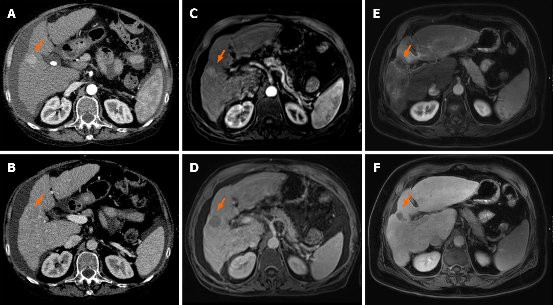
Figure 5 Imaging from before and after combination therapy in one patient in the transarterial chemoembolization + stereotactic body radiation therapy cohort.
A, B: Contrast enhanced computed tomography (CT); C-F: Magnetic resonance imaging (MRI). Contrast enhanced CT and MRI, arterial phase (top row) and portal venous phase (bottom row) cross sectional imaging from before (A–D) and after (E, F) treatment. At baseline CT and MRI, a well-defined nodular lesion with typical contrast agent dynamics is noted in the right liver lobe (Arrows). After treatment, typical radiation induced peri-lesional hyperenhancement and no hepatocellular carcinoma-specific contrast agent uptake is noted.
- Citation: Bauer U, Gerum S, Roeder F, Münch S, Combs SE, Philipp AB, De Toni EN, Kirstein MM, Vogel A, Mogler C, Haller B, Neumann J, Braren RF, Makowski MR, Paprottka P, Guba M, Geisler F, Schmid RM, Umgelter A, Ehmer U. High rate of complete histopathological response in hepatocellular carcinoma patients after combined transarterial chemoembolization and stereotactic body radiation therapy. World J Gastroenterol 2021; 27(24): 3630-3642
- URL: https://www.wjgnet.com/1007-9327/full/v27/i24/3630.htm
- DOI: https://dx.doi.org/10.3748/wjg.v27.i24.3630


