Copyright
©The Author(s) 2021.
World J Gastroenterol. Jun 28, 2021; 27(24): 3630-3642
Published online Jun 28, 2021. doi: 10.3748/wjg.v27.i24.3630
Published online Jun 28, 2021. doi: 10.3748/wjg.v27.i24.3630
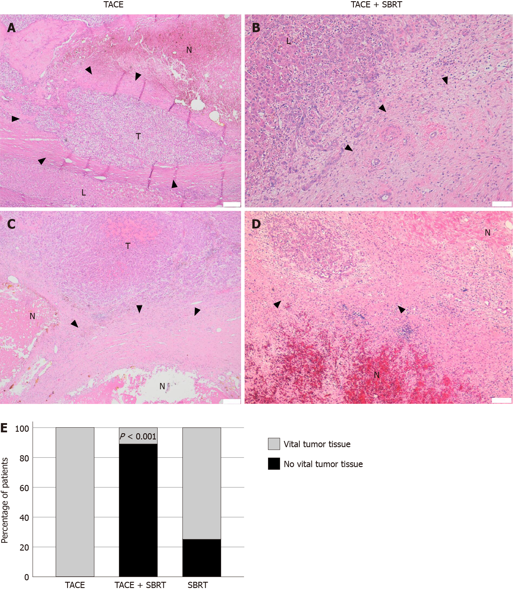
Figure 2 Tumor response by histopathology for each treatment group.
A-D: Representative histopathology (Hematoxylin and eosin stain) of tumor lesions in explant livers after transarterial chemoembolization (TACE) (A, C; scale bar 200 µm) or TACE + stereotactic body radiation therapy (SBRT) (B, D; scale bar 100 µm). Samples show necrosis with granulation tissue and organization by connective tissue at the border area (arrowheads) to normal liver. Residual tumor tissue was observed in TACE only samples, while no vital tumor cells could be detected in most patients in the TACE + SBRT group (B, D); E: Bar graph displaying the proportion of vital tumor tissue in each treatment group. Combination therapy with TACE and SBRT leads to a statistically significantly lower number of residual tumor tissue in explant livers (P < 0.001). TACE: Transarterial chemoembolization; SBRT: Stereotactic body radiation therapy; N: Necrosis; L: Normal liver; T: Tumor tissue.
- Citation: Bauer U, Gerum S, Roeder F, Münch S, Combs SE, Philipp AB, De Toni EN, Kirstein MM, Vogel A, Mogler C, Haller B, Neumann J, Braren RF, Makowski MR, Paprottka P, Guba M, Geisler F, Schmid RM, Umgelter A, Ehmer U. High rate of complete histopathological response in hepatocellular carcinoma patients after combined transarterial chemoembolization and stereotactic body radiation therapy. World J Gastroenterol 2021; 27(24): 3630-3642
- URL: https://www.wjgnet.com/1007-9327/full/v27/i24/3630.htm
- DOI: https://dx.doi.org/10.3748/wjg.v27.i24.3630


