Copyright
©The Author(s) 2021.
World J Gastroenterol. Jun 21, 2021; 27(23): 3327-3341
Published online Jun 21, 2021. doi: 10.3748/wjg.v27.i23.3327
Published online Jun 21, 2021. doi: 10.3748/wjg.v27.i23.3327
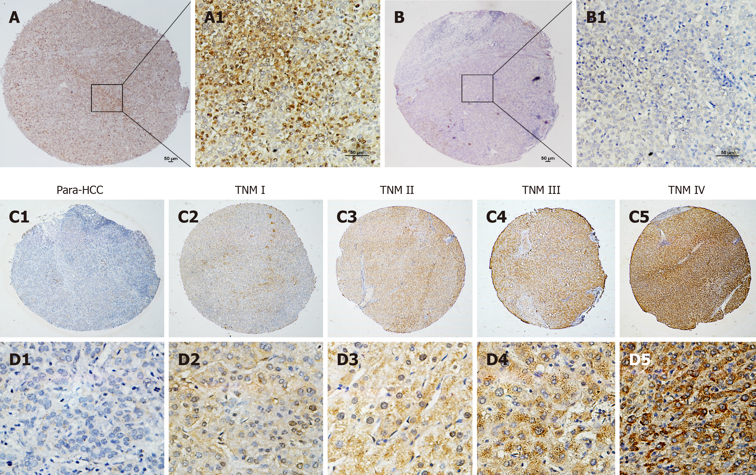
Figure 2 Tuftelin 1 expression with clinical staging of hepatocellular carcinoma.
A, A1: Hepatocellular carcinoma (HCC) tissue with strongly positive staining; B, B1: Adjacent non-HCC tissue with negative staining, original magnifications of × 40 (scale bar: 50 μm) in A and B, and × 200 (scale bar: 50 μm) in A1 and B1; C, D: Tuftelin 1 (TUFT1) expression in different HCC staging; C1, D1: Low TUFT1 expression in the non-HCC tissues; C2-C5: The brown staining of TUFT1 gradually increases in the HCC tissues from stage I to IV (original magnification × 40); D2-D5: The staining of TUFT1 in the non-HCC tissues from stage I to IV (original magnification × 400). TNM: Tumor-node-metastasis.
- Citation: Wu MN, Zheng WJ, Ye WX, Wang L, Chen Y, Yang J, Yao DF, Yao M. Oncogenic tuftelin 1 as a potential molecular-targeted for inhibiting hepatocellular carcinoma growth. World J Gastroenterol 2021; 27(23): 3327-3341
- URL: https://www.wjgnet.com/1007-9327/full/v27/i23/3327.htm
- DOI: https://dx.doi.org/10.3748/wjg.v27.i23.3327


