Copyright
©The Author(s) 2021.
World J Gastroenterol. Jun 7, 2021; 27(21): 2895-2909
Published online Jun 7, 2021. doi: 10.3748/wjg.v27.i21.2895
Published online Jun 7, 2021. doi: 10.3748/wjg.v27.i21.2895
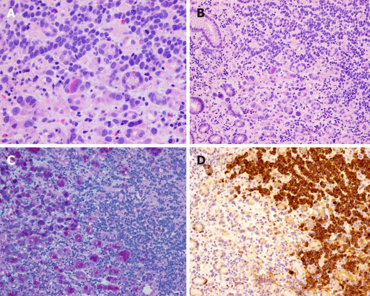
Figure 4 Morphology and immunohistochemical staining of mixed adenoneuroendocrine carcinoma.
A and B: Morphology of gastric small cell neuroendocrine carcinoma mixed with adenocarcinoma (hematoxylin and eosin staining, × 400); C: Gastric small cell neuroendocrine carcinoma mixed with adenocarcinoma (hematoxylin and eosin staining, × 200). Alcian blue/periodic acid–Schiff (AB-PAS) staining (× 200): The left side of the picture shows adenocarcinoma (AB-PAS positive); D: Chromogranin A-positive staining (× 200). The right side of the picture shows small cell neuroendocrine carcinoma.
- Citation: Han D, Li YL, Zhou ZW, Yin F, Chen J, Liu F, Shi YF, Wang W, Zhang Y, Yu XJ, Xu JM, Yang RX, Tian C, Luo J, Tan HY. Clinicopathological characteristics and prognosis of 232 patients with poorly differentiated gastric neuroendocrine neoplasms. World J Gastroenterol 2021; 27(21): 2895-2909
- URL: https://www.wjgnet.com/1007-9327/full/v27/i21/2895.htm
- DOI: https://dx.doi.org/10.3748/wjg.v27.i21.2895


