Copyright
©The Author(s) 2021.
World J Gastroenterol. Jun 7, 2021; 27(21): 2895-2909
Published online Jun 7, 2021. doi: 10.3748/wjg.v27.i21.2895
Published online Jun 7, 2021. doi: 10.3748/wjg.v27.i21.2895
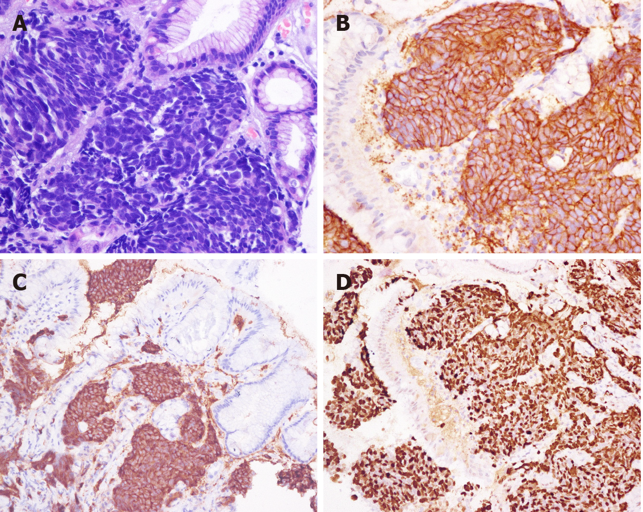
Figure 3 Morphology and immunohistochemical staining of gastric small cell neuroendocrine carcinoma.
A: Morphology (hematoxylin and eosin staining, × 400); B: CD56-positive staining [immunohistochemical (IHC), × 400]; C: Synaptophysin-positive staining (IHC, × 200); D: Ki-67 index: 90% (IHC, × 200).
- Citation: Han D, Li YL, Zhou ZW, Yin F, Chen J, Liu F, Shi YF, Wang W, Zhang Y, Yu XJ, Xu JM, Yang RX, Tian C, Luo J, Tan HY. Clinicopathological characteristics and prognosis of 232 patients with poorly differentiated gastric neuroendocrine neoplasms. World J Gastroenterol 2021; 27(21): 2895-2909
- URL: https://www.wjgnet.com/1007-9327/full/v27/i21/2895.htm
- DOI: https://dx.doi.org/10.3748/wjg.v27.i21.2895


