Copyright
©The Author(s) 2021.
World J Gastroenterol. Jun 7, 2021; 27(21): 2895-2909
Published online Jun 7, 2021. doi: 10.3748/wjg.v27.i21.2895
Published online Jun 7, 2021. doi: 10.3748/wjg.v27.i21.2895
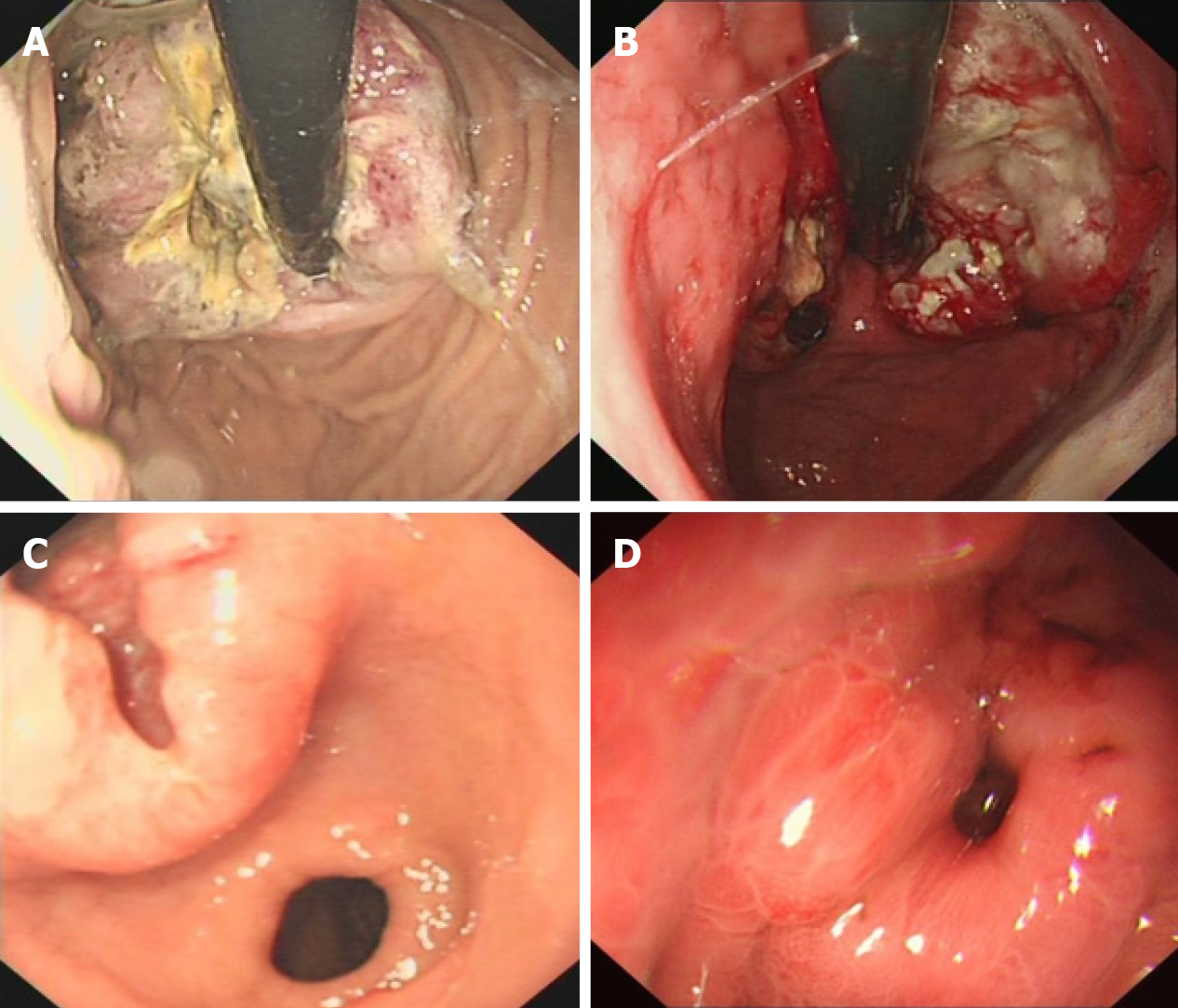
Figure 1 Endoscopic detection of poorly differentiated gastric neuroendocrine neoplasms.
A: Circumferential raised lesions on the cardia with uneven surfaces; B: Irregular bumps on the side of the minor curvature of the cardia, accompanied by erosions, ulcers, and unclear boundaries that bled easily when contacted; C: A raised ulcer with a diameter of 4 cm was observed in the small curvature of the antrum; D: A deep ulcer with a diameter of approximately 0.5 cm on the posterior wall of the gastric fundus was observed, and the base was not clear.
- Citation: Han D, Li YL, Zhou ZW, Yin F, Chen J, Liu F, Shi YF, Wang W, Zhang Y, Yu XJ, Xu JM, Yang RX, Tian C, Luo J, Tan HY. Clinicopathological characteristics and prognosis of 232 patients with poorly differentiated gastric neuroendocrine neoplasms. World J Gastroenterol 2021; 27(21): 2895-2909
- URL: https://www.wjgnet.com/1007-9327/full/v27/i21/2895.htm
- DOI: https://dx.doi.org/10.3748/wjg.v27.i21.2895


