Copyright
©The Author(s) 2021.
World J Gastroenterol. May 28, 2021; 27(20): 2545-2575
Published online May 28, 2021. doi: 10.3748/wjg.v27.i20.2545
Published online May 28, 2021. doi: 10.3748/wjg.v27.i20.2545
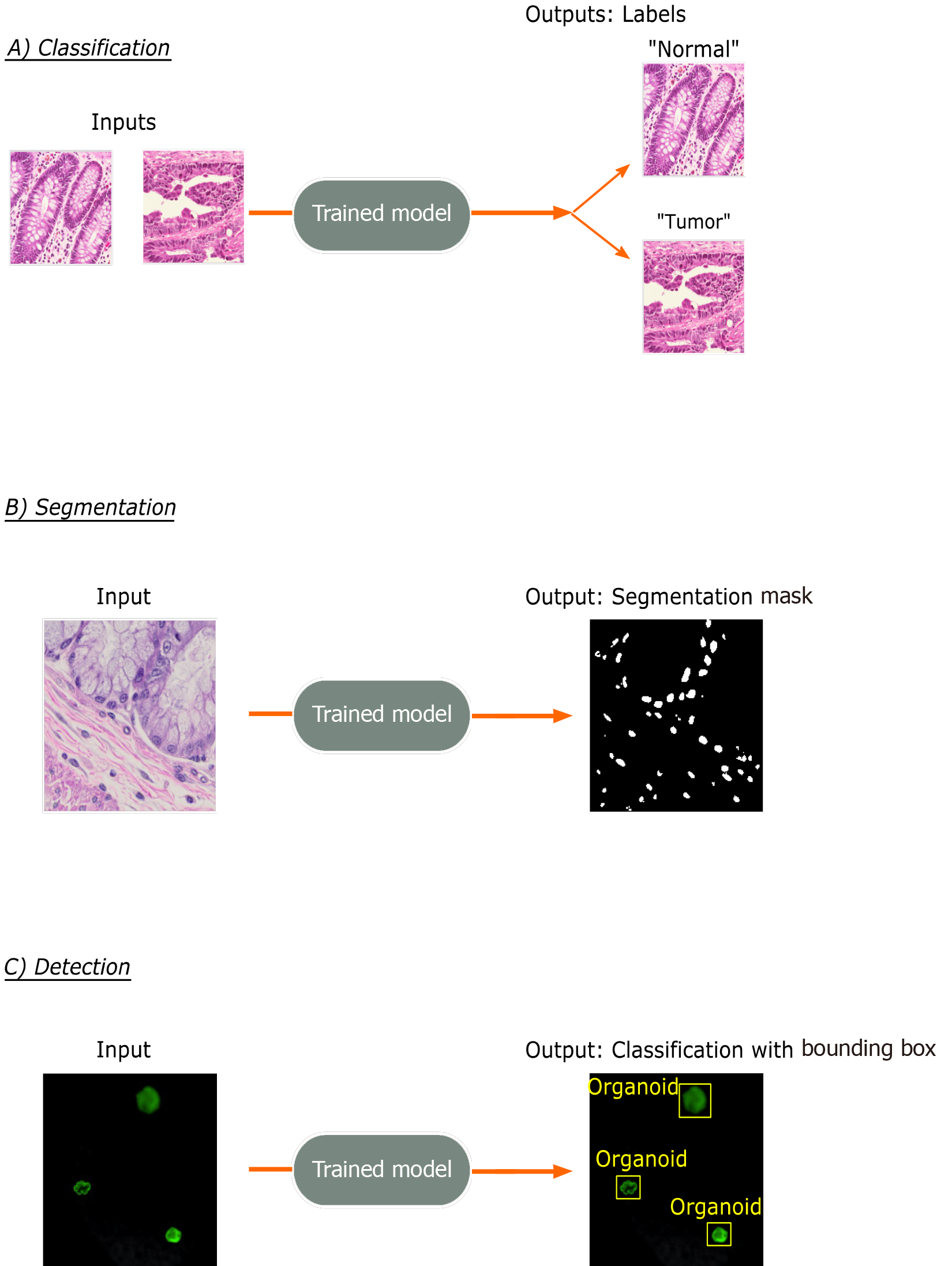
Figure 2 Common trainable tasks by deep learning.
A: Classification involves designation of a class label to an image input. Image patches for the figure were taken from colorectal cancer and normal adjacent intestinal samples obtained via an IRB-approved protocol; B: Segmentation tasks output a mask with pixel-level color designation of classes. Here, white indicates nuclei and black represents non-nuclear areas; C: Detection tasks generate bounding boxes with object classifications. Immunofluorescence images of mouse-derived organoids with manually inserted classifications and bounding boxes in yellow are included for illustrative purposes.
- Citation: Kobayashi S, Saltz JH, Yang VW. State of machine and deep learning in histopathological applications in digestive diseases. World J Gastroenterol 2021; 27(20): 2545-2575
- URL: https://www.wjgnet.com/1007-9327/full/v27/i20/2545.htm
- DOI: https://dx.doi.org/10.3748/wjg.v27.i20.2545


