Copyright
©The Author(s) 2021.
World J Gastroenterol. Apr 21, 2021; 27(15): 1569-1577
Published online Apr 21, 2021. doi: 10.3748/wjg.v27.i15.1569
Published online Apr 21, 2021. doi: 10.3748/wjg.v27.i15.1569
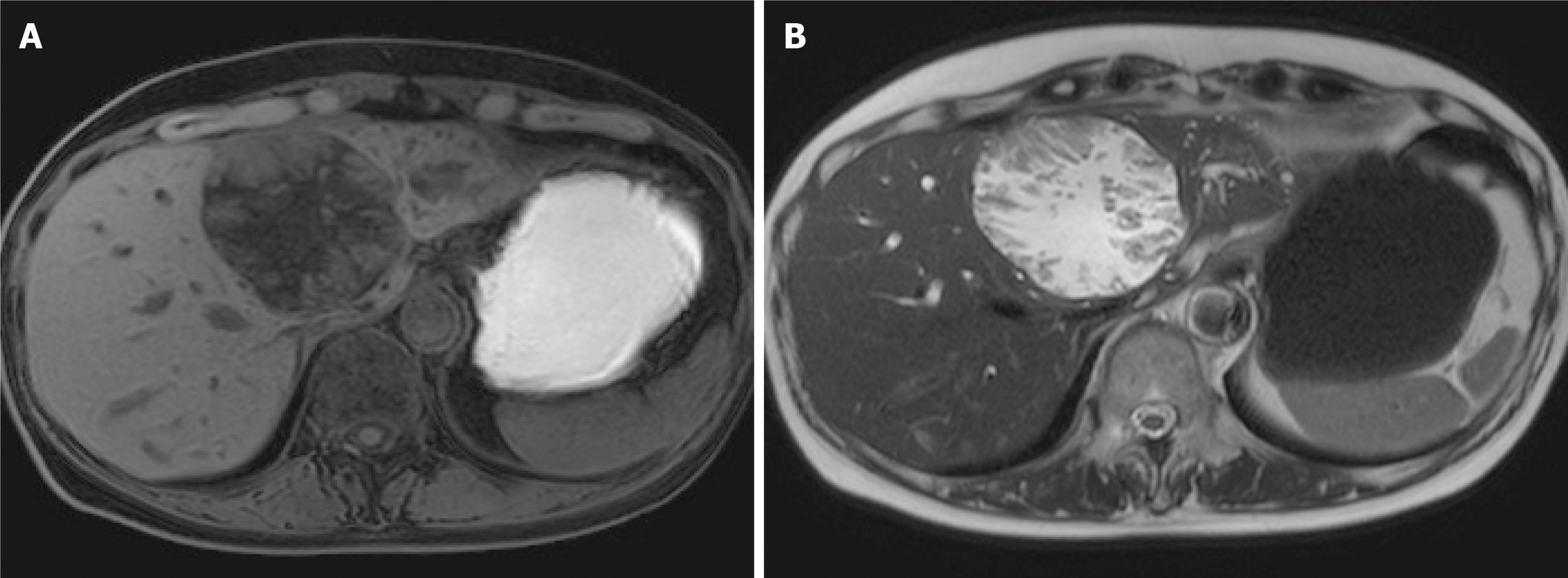
Figure 2 Magnetic resonance imaging.
A: T1-weighted image; and B: T2-weighted image papillary solid lesion protuberating from cystic wall is noted and the cystic component showed the same signals as water. Transportation between the cyst and the root of left hepatic duct is noted.
- Citation: Sakai Y, Ohtsuka M, Sugiyama H, Mikata R, Yasui S, Ohno I, Iino Y, Kato J, Tsuyuguchi T, Kato N. Current status of diagnosis and therapy for intraductal papillary neoplasm of the bile duct. World J Gastroenterol 2021; 27(15): 1569-1577
- URL: https://www.wjgnet.com/1007-9327/full/v27/i15/1569.htm
- DOI: https://dx.doi.org/10.3748/wjg.v27.i15.1569


