Copyright
©The Author(s) 2021.
World J Gastroenterol. Apr 14, 2021; 27(14): 1435-1450
Published online Apr 14, 2021. doi: 10.3748/wjg.v27.i14.1435
Published online Apr 14, 2021. doi: 10.3748/wjg.v27.i14.1435
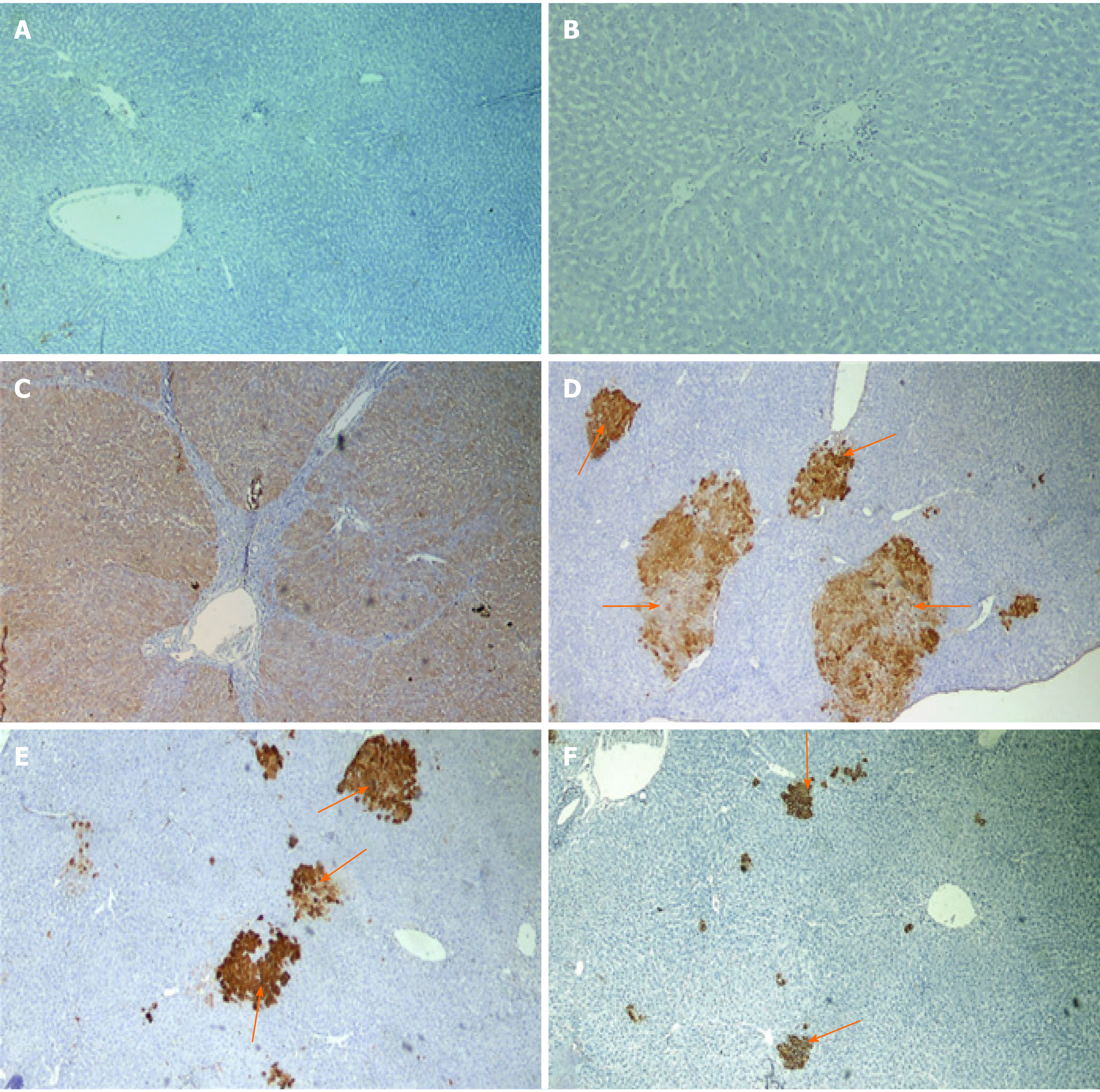
Figure 5 Photomicrographs of liver sections of rats immunohistochemical stained with glutathione S-transferase placental antibody.
A and B: Naive group; C: Precancerous lesion group showing multiple glutathione S-transferase placental (GSTP)-positive large hepatic nodules (brown stained nodules) occupying most of section; D-F: Liver sections of rats treated with different doses of cyanidin (10, 15, 20 mg/kg) showing GSTP positive small hepatic foci (brown stained cells = arrow) of different size scattered in-between negatively stained hepatic parenchyma. (Magnification: × 40)
- Citation: Matboli M, Hasanin AH, Hussein R, El-Nakeep S, Habib EK, Ellackany R, Saleh LA. Cyanidin 3-glucoside modulated cell cycle progression in liver precancerous lesion, in vivo study. World J Gastroenterol 2021; 27(14): 1435-1450
- URL: https://www.wjgnet.com/1007-9327/full/v27/i14/1435.htm
- DOI: https://dx.doi.org/10.3748/wjg.v27.i14.1435


