Copyright
©The Author(s) 2021.
World J Gastroenterol. Apr 14, 2021; 27(14): 1435-1450
Published online Apr 14, 2021. doi: 10.3748/wjg.v27.i14.1435
Published online Apr 14, 2021. doi: 10.3748/wjg.v27.i14.1435
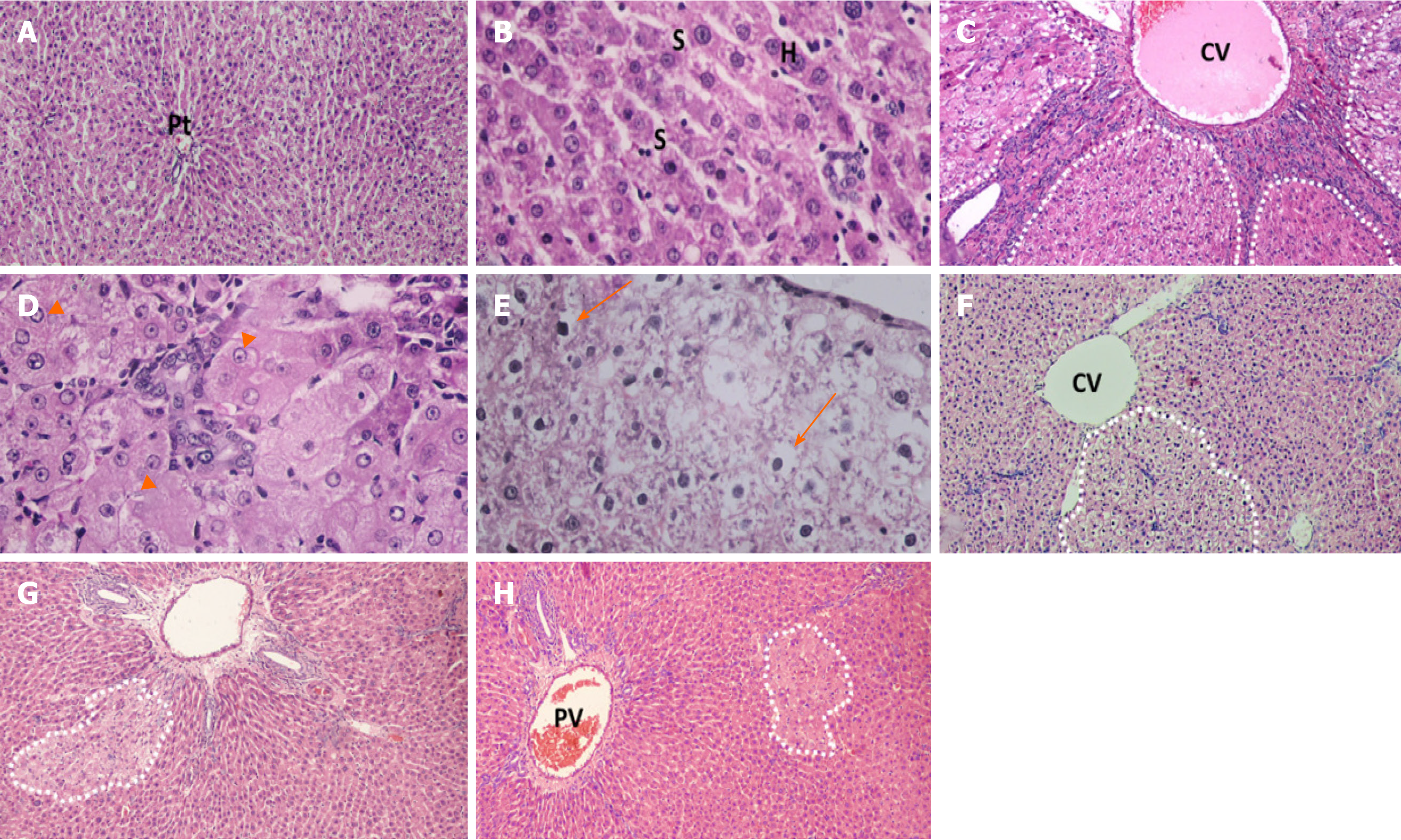
Figure 4 Photomicrographs of liver sections stained with H&E staining.
A and B: Naïve group liver sections showed normal hepatic architecture, cords of hepatocytes radiating from central vein and portal triads present in-between and polygonal hepatocytes with central rounded vesicular nuclei, and hepatic sinusoid in-between; C-E: Liver sections of rats received diethylnitrosamine/2-acetylaminofluorene (DEN/2-AAF) showed larger, discriminated dysplastic nodules (doted shapes) compressing the surrounding liver tissue with disruption of normal hepatic lobular architecture; D: Liver sections of rats received DEN/2-AAF showed eosinophilic foci of cellular alteration consisting of enlarged hepatocytes with increased acidophilic staining and vaculated nuclei (arrow head); E: Liver sections of rats received DEN/2-AAF showed foci of Clear cell formed of hepatocytes showing variable degrees of cytoplasmic vacuolations and ballooning with pyknotic nuclei (arrow); F-H: Liver sections of rats treated with different doses of cyanidin (10, 15, 20 mg/kg) respectively, showing small and less discriminated dysplastic nodules (doted shapes). (Magnification: A, C, F, G, H × 1000; B, D, E × 400). CV: Central vein; PV: Portal vein; Pt: Portal triads; S: Sinusoid; H: Hepatocytes.
- Citation: Matboli M, Hasanin AH, Hussein R, El-Nakeep S, Habib EK, Ellackany R, Saleh LA. Cyanidin 3-glucoside modulated cell cycle progression in liver precancerous lesion, in vivo study. World J Gastroenterol 2021; 27(14): 1435-1450
- URL: https://www.wjgnet.com/1007-9327/full/v27/i14/1435.htm
- DOI: https://dx.doi.org/10.3748/wjg.v27.i14.1435


