Copyright
©The Author(s) 2021.
World J Gastroenterol. Apr 14, 2021; 27(14): 1419-1434
Published online Apr 14, 2021. doi: 10.3748/wjg.v27.i14.1419
Published online Apr 14, 2021. doi: 10.3748/wjg.v27.i14.1419
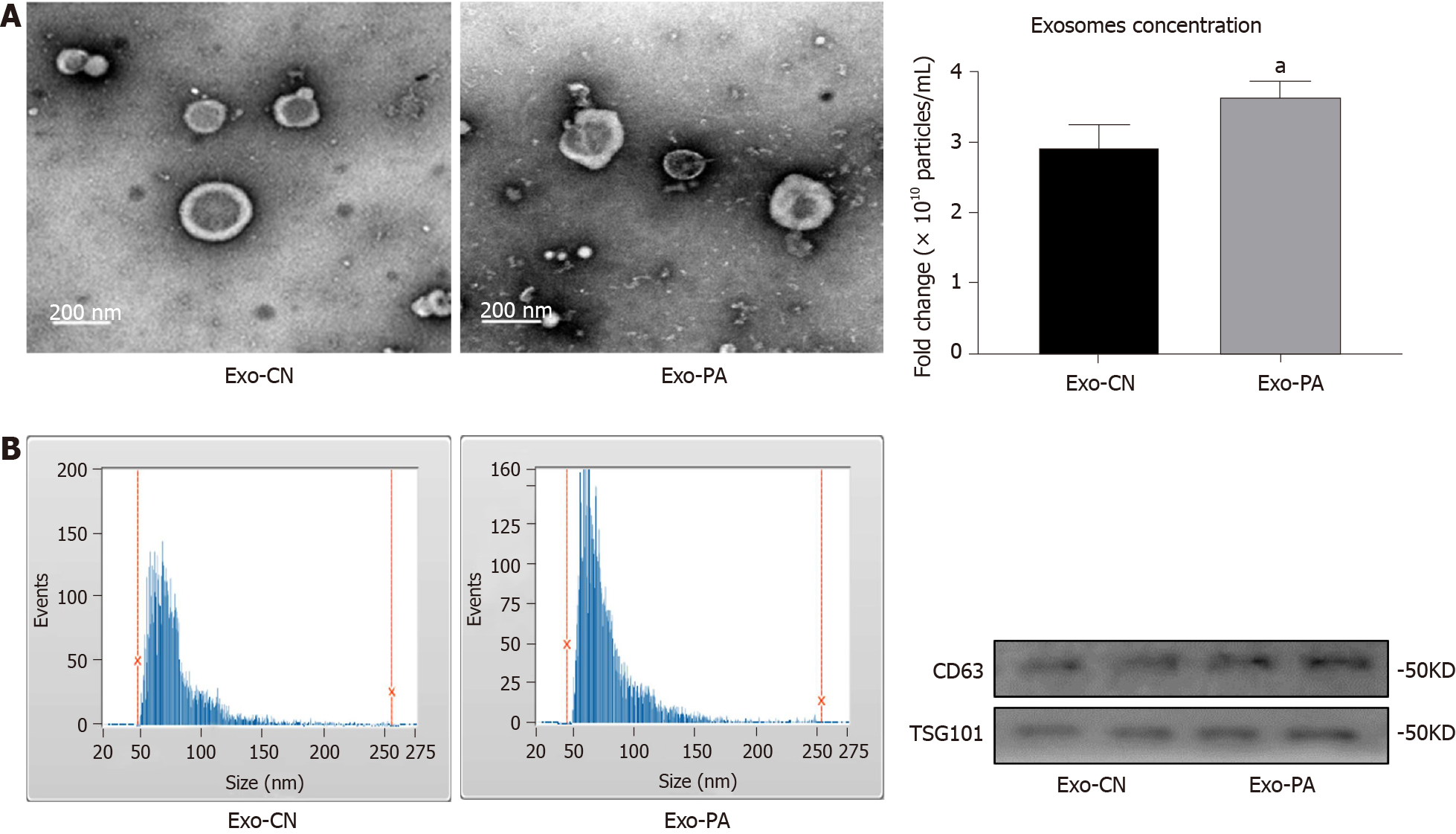
Figure 1 Identification of exosomes derived from primary hepatocytes.
A: Exosomes derived from primary hepatocytes (Exo-PHC) were visualized by electronic microscopy (× 30000); B: Nanoparticle tracking analysis for Exo-PHC; C: The concentration of Exo-PHC was measured. Exosomes derived from vehicle control (Exo-CN) treated PHC: 2.92 ± 0.32 × 10¹⁰ particles/mL vs exosomes derived from palmitic acid (Exo-PA) treated PHC: 3.62 ± 0.22 × 10¹⁰ particles/mL; D: Exosomal markers CD63 and TSG101 were detected by western blot in Exo-PHC. Statistical significance, aP < 0.05.
- Citation: Luo X, Luo SZ, Xu ZX, Zhou C, Li ZH, Zhou XY, Xu MY. Lipotoxic hepatocyte-derived exosomal miR-1297 promotes hepatic stellate cell activation through the PTEN signaling pathway in metabolic-associated fatty liver disease. World J Gastroenterol 2021; 27(14): 1419-1434
- URL: https://www.wjgnet.com/1007-9327/full/v27/i14/1419.htm
- DOI: https://dx.doi.org/10.3748/wjg.v27.i14.1419


