Copyright
©The Author(s) 2021.
World J Gastroenterol. Apr 7, 2021; 27(13): 1354-1361
Published online Apr 7, 2021. doi: 10.3748/wjg.v27.i13.1354
Published online Apr 7, 2021. doi: 10.3748/wjg.v27.i13.1354
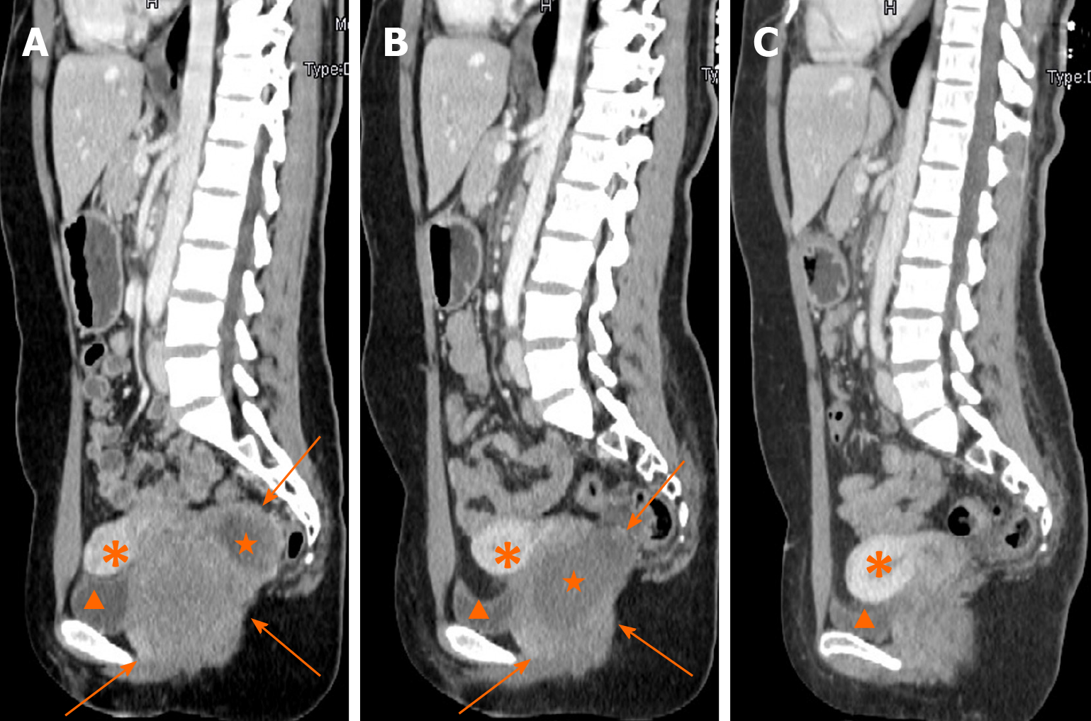
Figure 1 Contrast-enhanced computed tomography in sagittal section.
A: A rectovaginal space mass of 8.0 cm in maximum diameter with non-enhancing liquefaction necrosis at the time of admission; B: The mass significantly shrunk to 6.4 cm in maximum diameter after 5 mo of imatinib treatment and non-enhanced liquefaction area enlarged clearly; C: No obvious sign of tumor recurrence in rectovaginal space 15 mo after tumor resection. Arrows: The mass; pentagrams: Liquefaction necrosis area; arrow heads: Bladder; asterisks: Uterus.
- Citation: Zhang Q, Zhao JY, Zhuang H, Lu CY, Yao J, Luo Y, Yu YY. Transperineal core-needle biopsy of a rectal subepithelial lesion guided by endorectal ultrasound after contrast-enhanced ultrasound: A case report. World J Gastroenterol 2021; 27(13): 1354-1361
- URL: https://www.wjgnet.com/1007-9327/full/v27/i13/1354.htm
- DOI: https://dx.doi.org/10.3748/wjg.v27.i13.1354


