Copyright
©The Author(s) 2021.
World J Gastroenterol. Jan 7, 2021; 27(1): 55-68
Published online Jan 7, 2021. doi: 10.3748/wjg.v27.i1.55
Published online Jan 7, 2021. doi: 10.3748/wjg.v27.i1.55
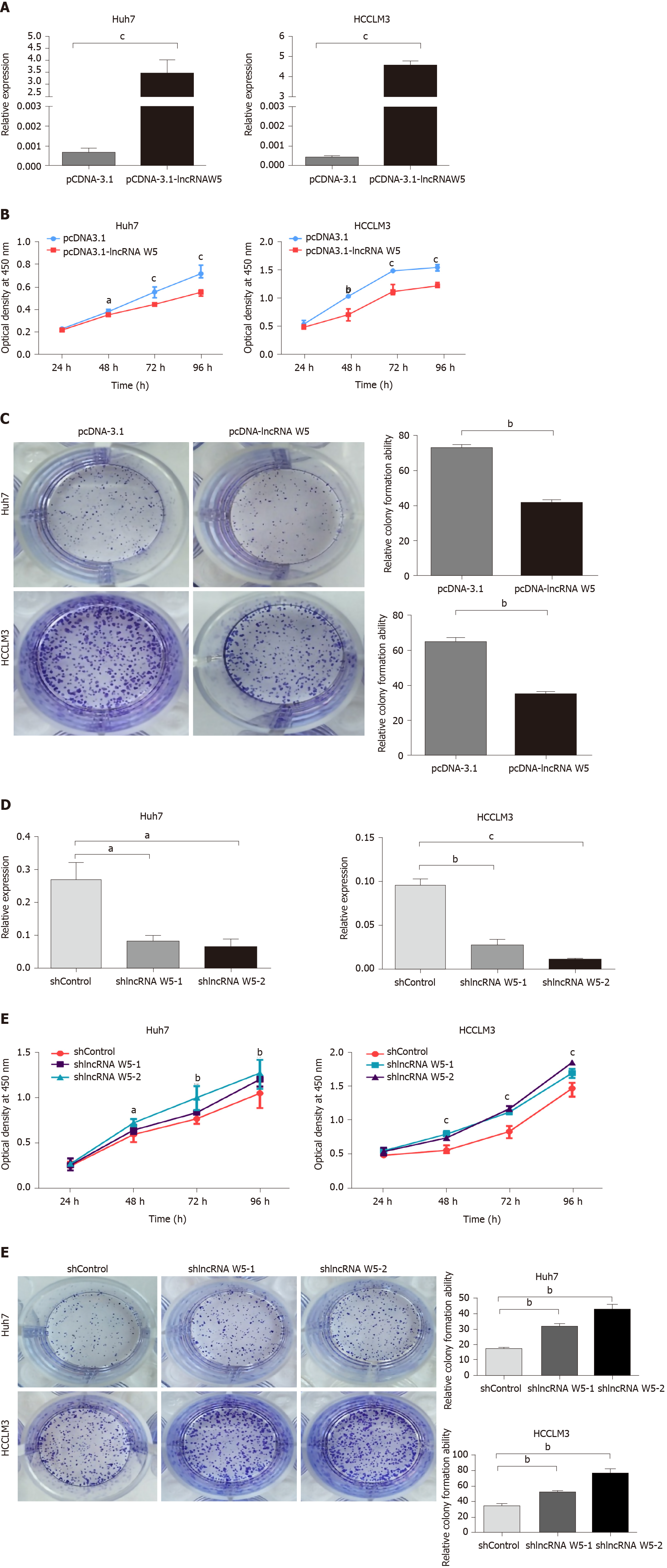
Figure 2 In vitro suppression of long non-coding ribonucleic acid W5 in hepatocellular carcinoma proliferation.
A: Increased long non-coding ribonucleic acid (lncRNA) W5 expression in Huh7 and LM3 cells was confirmed after over-expressed lncRNA W5 transfection by reverse transcription-polymerase chain reaction. LncRNA W5 expression was normalized to GAPDH. cP < 0.001; B: Cell viability of pCDNA-3.1 LncRNAW5-transfected Huh7 and LM3 cells were detected by CCK-8 assays. Cell number was determined every 24 h up to 96 h. The results are shown as the mean ± SE from three independent experiments. aP < 0.05; bP < 0.01. cP < 0.001, compared with the control by two-sided t-test; C: Colony-forming assay was used to determine the effect of lncRNA W5 on the proliferation in Huh7 and LM3 cells; D: Decreased lncRNA W5 expression in Huh7 and LM3 cells was confirmed after sh-1 or sh-2 LncRNAW5 transfection by reverse transcription-polymerase chain reaction. LncRNA W5 expression was normalized to GAPDH. aP < 0.05; bP < 0.01. cP < 0.001; E: Cell viability of sh-1 or sh-2 LncRNAW5-transfected Huh7 and LM3 cells were detected by CCK-8 assays. Cell number was determined every 24 h up to 96 h. The results are shown as the mean ± SE from at least three independent experiments. aP < 0.05; bP < 0.01. cP < 0.001, compared with the control by two-sided t-test; and F: Colony-forming assay was used to determine the effect of sh-1 or sh-2 LncRNA W5 on the proliferation of Huh7 and LM3 cells.
- Citation: Lei GL, Fan HX, Wang C, Niu Y, Li TL, Yu LX, Hong ZX, Yan J, Wang XL, Zhang SG, Ren MJ, Yang PH. Long non-coding ribonucleic acid W5 inhibits progression and predicts favorable prognosis in hepatocellular carcinoma. World J Gastroenterol 2021; 27(1): 55-68
- URL: https://www.wjgnet.com/1007-9327/full/v27/i1/55.htm
- DOI: https://dx.doi.org/10.3748/wjg.v27.i1.55


