Copyright
©The Author(s) 2021.
World J Gastroenterol. Jan 7, 2021; 27(1): 129-142
Published online Jan 7, 2021. doi: 10.3748/wjg.v27.i1.129
Published online Jan 7, 2021. doi: 10.3748/wjg.v27.i1.129
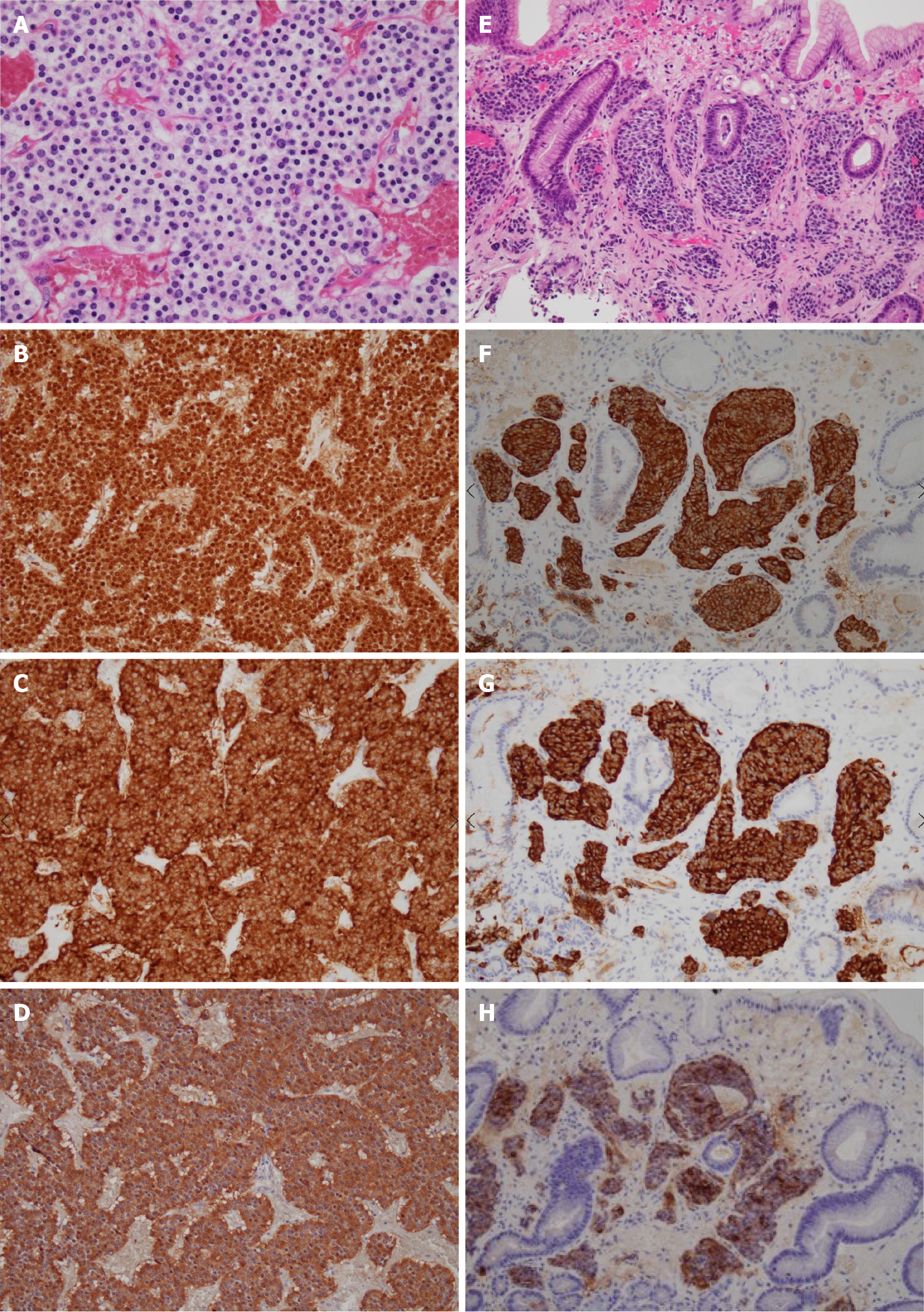
Figure 4 Pathology of the surgical specimen (A-D) and gastric biopsy (E-H).
Nests of tumor cells characterized by small ovoid nuclei and mildly eosinophilic cytoplasms with intervening dilated capillary networks were observed in the omental lesion (A). The tumor was positive for chromogranin A (B), synaptophysin (C), and gastrin (D) stains. Biopsy of the gastric lesion showed similar cells in the mucosal layer (E) which were also positive for chromogranin A (F), synaptophysin (G), and gastrin (H) stains.
- Citation: Okamoto T, Yoshimoto T, Ohike N, Fujikawa A, Kanie T, Fukuda K. Spontaneous regression of gastric gastrinoma after resection of metastases to the lesser omentum: A case report and review of literature. World J Gastroenterol 2021; 27(1): 129-142
- URL: https://www.wjgnet.com/1007-9327/full/v27/i1/129.htm
- DOI: https://dx.doi.org/10.3748/wjg.v27.i1.129


