Copyright
©The Author(s) 2021.
World J Gastroenterol. Jan 7, 2021; 27(1): 129-142
Published online Jan 7, 2021. doi: 10.3748/wjg.v27.i1.129
Published online Jan 7, 2021. doi: 10.3748/wjg.v27.i1.129
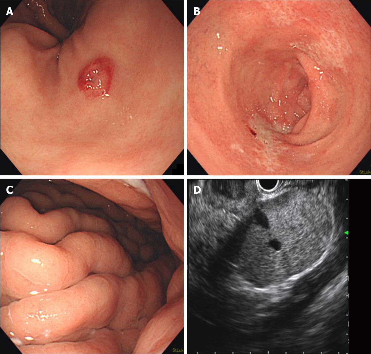
Figure 2 Esophagogastroduodenoscopy incidentally revealed a red, 7 mm submucosal tumor with a central depression (A); prominent gastric folds (B) and shallow duodenal ulcers (C) were also noted; endoscopic ultrasound revealed 3 clearly delineated, hyperechoic masses with uniform texture, of which 1 had a hypoechoic center with clear borders (D).
- Citation: Okamoto T, Yoshimoto T, Ohike N, Fujikawa A, Kanie T, Fukuda K. Spontaneous regression of gastric gastrinoma after resection of metastases to the lesser omentum: A case report and review of literature. World J Gastroenterol 2021; 27(1): 129-142
- URL: https://www.wjgnet.com/1007-9327/full/v27/i1/129.htm
- DOI: https://dx.doi.org/10.3748/wjg.v27.i1.129


