Copyright
©The Author(s) 2020.
World J Gastroenterol. Mar 7, 2020; 26(9): 984-991
Published online Mar 7, 2020. doi: 10.3748/wjg.v26.i9.984
Published online Mar 7, 2020. doi: 10.3748/wjg.v26.i9.984
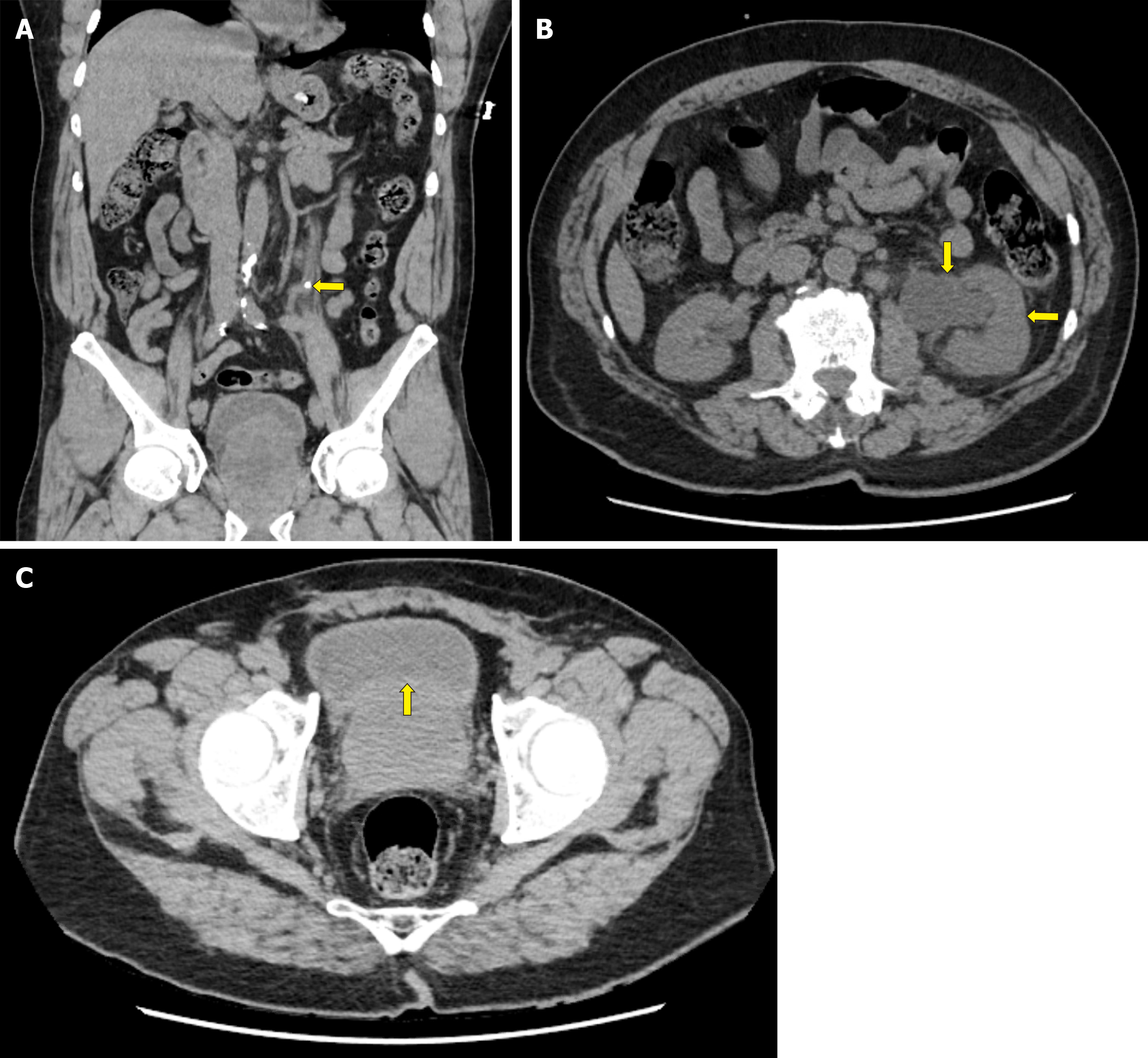
Figure 1 Abdomino-pelvic computed tomography without contrast.
A: Left kidney stone. Sagittal section of abdomino-pelvic computerized tomograph without IV contrast (not administered due to elevated creatinine) performed on admission in patient reported as case-1 demonstrates a 5.5-mm-wide, round, radiopaque, kidney stone in left ureter (arrow), just rostral to the level of left iliac crest; B: Left-sided hydroureteronephrosis. Axial section of the same abdomino-pelvic CT at level of mid-kidneys shows that this stone has caused left ureteral obstruction, left-sided hydroureter, and left-sided hydronephrosis. Note the severely dilated left renal calyx (vertical arrow) and compressed left renal parenchyma (horizontal arrow), as compared to normal-sized right calyx and right kidney; C: Moderately severe diffuse prostatomegaly. Axial section of the same abdomino-pelvic CT at level of rectum reveals moderately severe diffuse prostatomegaly, as demonstrated by the prostate compressing the bladder [arrow shows upper (ventral) margin of prostrate compressing bladder].
- Citation: Cappell MS. Two case reports of novel syndrome of bizarre performance of gastrointestinal endoscopy due to toxic encephalopathy of endoscopists among 181767 endoscopies in a 13-year-university hospital review: Endoscopists, first do no harm! World J Gastroenterol 2020; 26(9): 984-991
- URL: https://www.wjgnet.com/1007-9327/full/v26/i9/984.htm
- DOI: https://dx.doi.org/10.3748/wjg.v26.i9.984


