Copyright
©The Author(s) 2020.
World J Gastroenterol. Feb 14, 2020; 26(6): 686-695
Published online Feb 14, 2020. doi: 10.3748/wjg.v26.i6.686
Published online Feb 14, 2020. doi: 10.3748/wjg.v26.i6.686
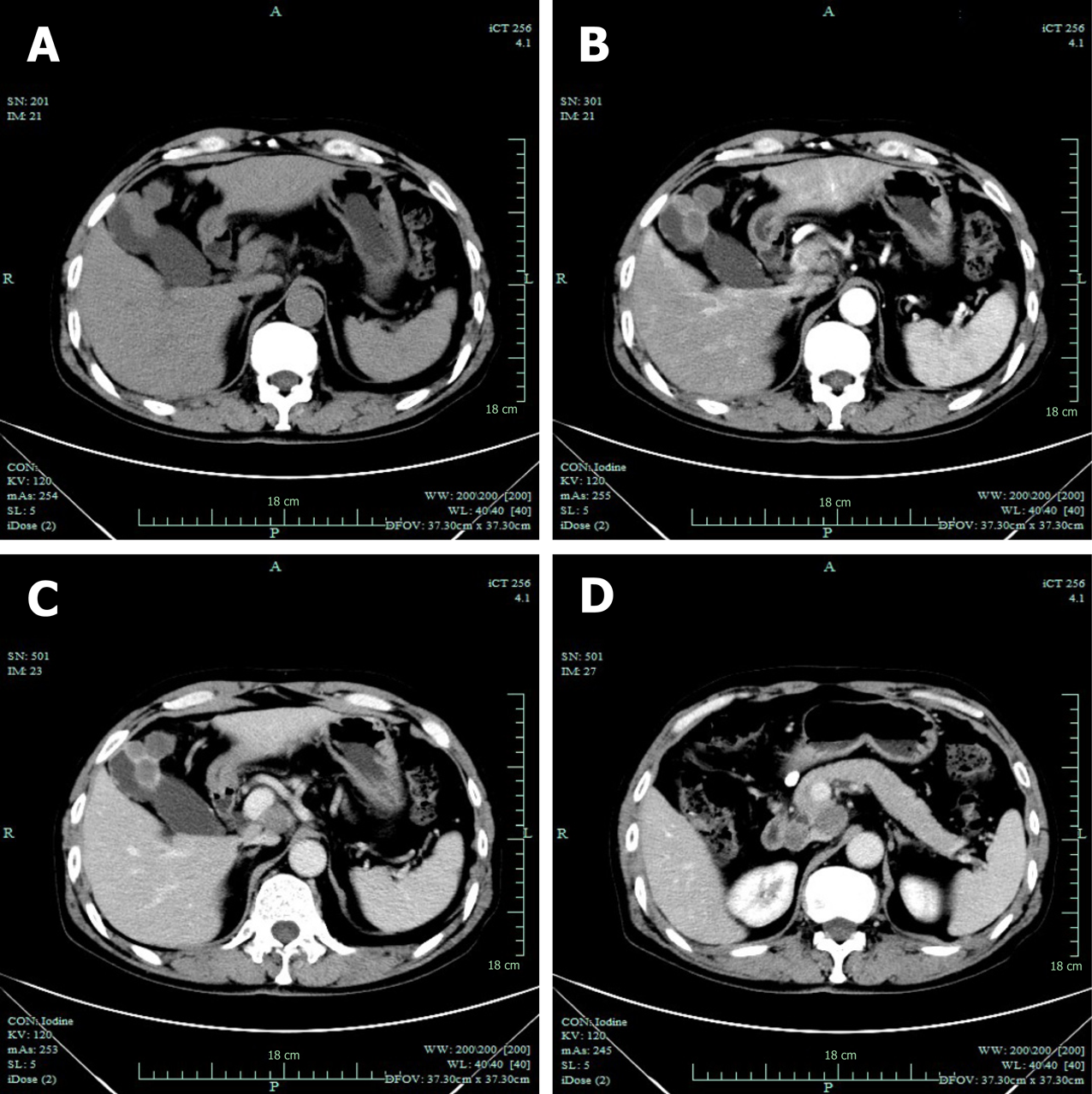
Figure 2 Computed tomography examination.
A: Gallbladder neoplasm was visible in unenhanced imagery; B: Gallbladder neoplasm was mildly enhanced in arterial phases; C: Gallbladder neoplasm was mildly enhanced in venous phases; D: Enlarged lymph nodes were seen in the portacaval space.
- Citation: Jin M, Zhou B, Jiang XL, Zhang QY, Zheng X, Jiang YC, Yan S. Flushing as atypical initial presentation of functional gallbladder neuroendocrine carcinoma: A case report. World J Gastroenterol 2020; 26(6): 686-695
- URL: https://www.wjgnet.com/1007-9327/full/v26/i6/686.htm
- DOI: https://dx.doi.org/10.3748/wjg.v26.i6.686


