Copyright
©The Author(s) 2020.
World J Gastroenterol. Dec 28, 2020; 26(48): 7707-7718
Published online Dec 28, 2020. doi: 10.3748/wjg.v26.i48.7707
Published online Dec 28, 2020. doi: 10.3748/wjg.v26.i48.7707
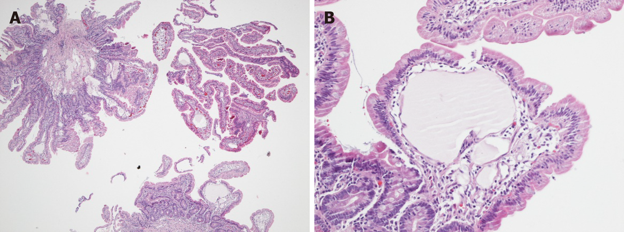
Figure 2 Histological and immunohistological analyses.
A: Jejunal biopsies showing a mild and focal blunting of the villi in particular above the prominent ecstatic mucosal lymph vessels (4-fold magnification); B: Ecstatic lymph vessel without inflammatory changes or abnormalities of the epithelial intestinal cell lining (200-fold magnification).
- Citation: Huber R, Semmler G, Mayr A, Offner F, Datz C. Primary intestinal lymphangiectasia in an adult patient: A case report and review of literature. World J Gastroenterol 2020; 26(48): 7707-7718
- URL: https://www.wjgnet.com/1007-9327/full/v26/i48/7707.htm
- DOI: https://dx.doi.org/10.3748/wjg.v26.i48.7707


