Copyright
©The Author(s) 2020.
World J Gastroenterol. Dec 21, 2020; 26(47): 7513-7527
Published online Dec 21, 2020. doi: 10.3748/wjg.v26.i47.7513
Published online Dec 21, 2020. doi: 10.3748/wjg.v26.i47.7513
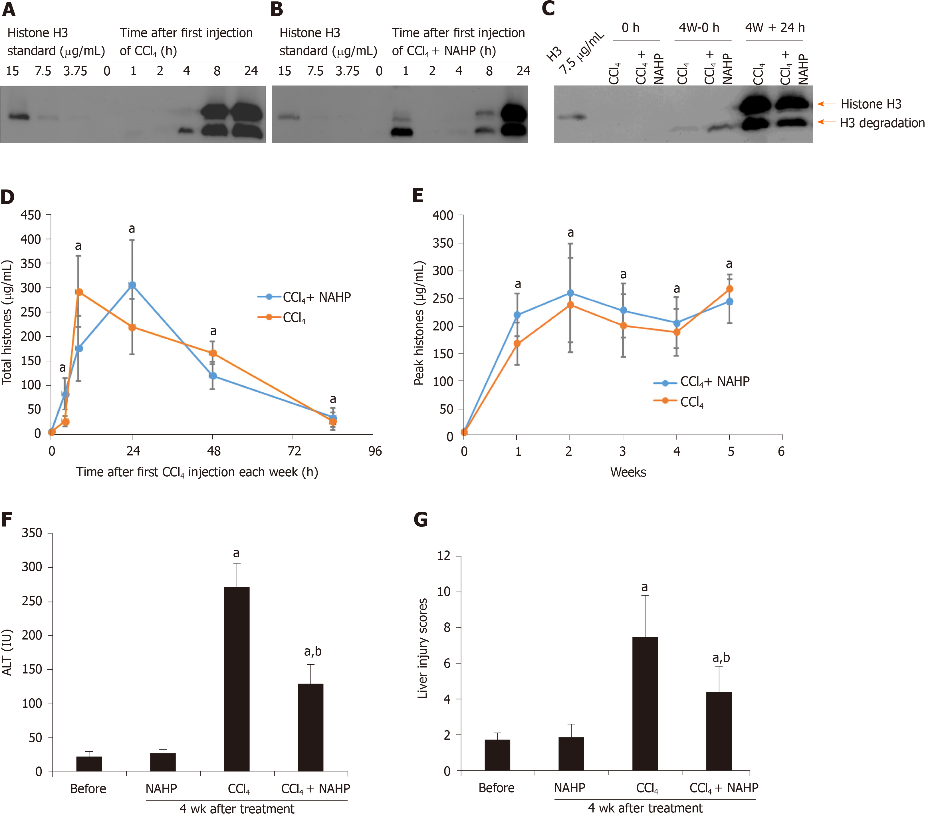
Figure 1 Circulating histones are elevated in ICR mice treated with CCl4.
Typical western blots of histone H3 standard and histone H3 in plasma from mice treated with the first dose of CCl4 (A) and CCl4 + non-anticoagulant heparin (NAHP) (B), and a typical H3 blot before and 24 h after the last dose of CCl4 (C); D: The mean ± SD of circulating histones at different time points, including 0 h (before the experiment), and 4, 8, 24, 48 and 84 h after first dose of CCl4 (orange) and CCl4 + NAHP (blue) of each week are presented. ANOVA test, aP < 0.05 compared to time 0; E: The mean ± SD of peak circulating histones (8 and 24 h after first CCl4 injection of each week). ANOVA test, aP < 0.05 compared to time 0 (before first injection). The mean ± SE of blood alanine aminotransferase levels (F) and liver injury scores (G) from nine mice in each group following 4 wk of treatment. ANOVA test, aP < 0.05 compared to mice without treatment (before). bP < 0.05 compared to CCl4 group. ANOVA: Analysis of variance; NAHP: Non-anticoagulant heparin; ALT: Alanine aminotransferase.
- Citation: Wang Z, Cheng ZX, Abrams ST, Lin ZQ, Yates E, Yu Q, Yu WP, Chen PS, Toh CH, Wang GZ. Extracellular histones stimulate collagen expression in vitro and promote liver fibrogenesis in a mouse model via the TLR4-MyD88 signaling pathway. World J Gastroenterol 2020; 26(47): 7513-7527
- URL: https://www.wjgnet.com/1007-9327/full/v26/i47/7513.htm
- DOI: https://dx.doi.org/10.3748/wjg.v26.i47.7513


