Copyright
©The Author(s) 2020.
World J Gastroenterol. Dec 14, 2020; 26(46): 7325-7337
Published online Dec 14, 2020. doi: 10.3748/wjg.v26.i46.7325
Published online Dec 14, 2020. doi: 10.3748/wjg.v26.i46.7325
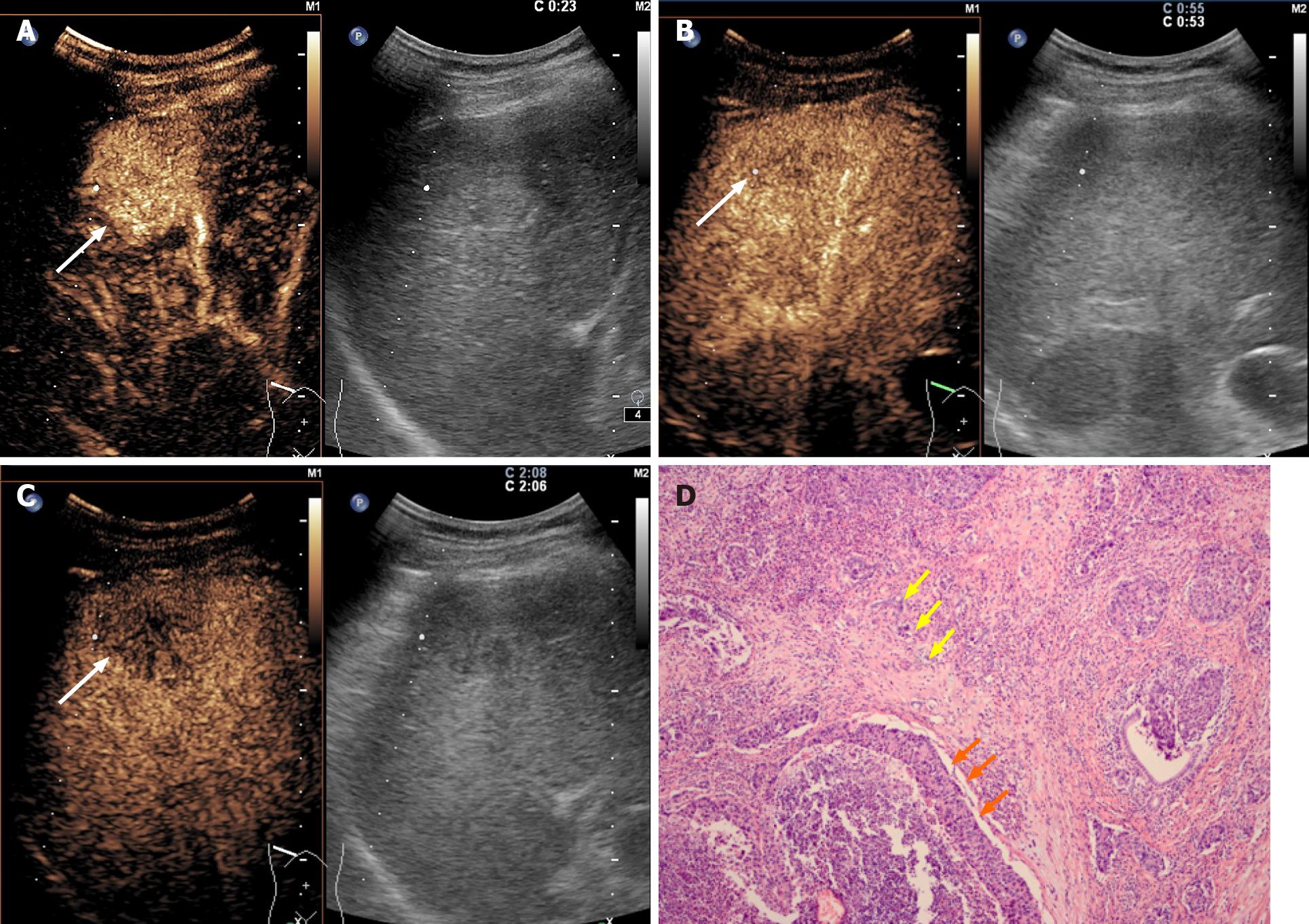
Figure 2 LR-M nodule in a 54-year-old man with chronic hepatitis B.
A: A nodule with a diameter of 3.6 cm in the right liver lobe was homogeneously hyper-enhanced (arrow) in the arterial phase at contrast-enhanced ultrasound; B: Early washout (53 s) of the contrast agent was observed (arrow); C: Hypo-enhancement (arrow) in the late phase was shown at contrast-enhanced ultrasonography. Elevated alpha-fetoprotein and normal carbohydrate antigen 19-9 level were found by in the serologic data. The nodule was assigned to combined hepatocellular-cholangiocarcinoma lesion according to the diagnostic criteria; D: Both hepatocellular carcinoma (orange arrow) and intrahepatic cholangiocarcinoma (yellow arrow) components were found in histopathologic analysis, resulting in a final diagnosis of combined hepatocellular-cholangiocarcinoma (hematoxylin and eosin staining; magnification, × 100).
- Citation: Yang J, Zhang YH, Li JW, Shi YY, Huang JY, Luo Y, Liu JB, Lu Q. Contrast-enhanced ultrasound in association with serum biomarkers for differentiating combined hepatocellular-cholangiocarcinoma from hepatocellular carcinoma and intrahepatic cholangiocarcinoma. World J Gastroenterol 2020; 26(46): 7325-7337
- URL: https://www.wjgnet.com/1007-9327/full/v26/i46/7325.htm
- DOI: https://dx.doi.org/10.3748/wjg.v26.i46.7325


