Copyright
©The Author(s) 2020.
World J Gastroenterol. Dec 14, 2020; 26(46): 7312-7324
Published online Dec 14, 2020. doi: 10.3748/wjg.v26.i46.7312
Published online Dec 14, 2020. doi: 10.3748/wjg.v26.i46.7312
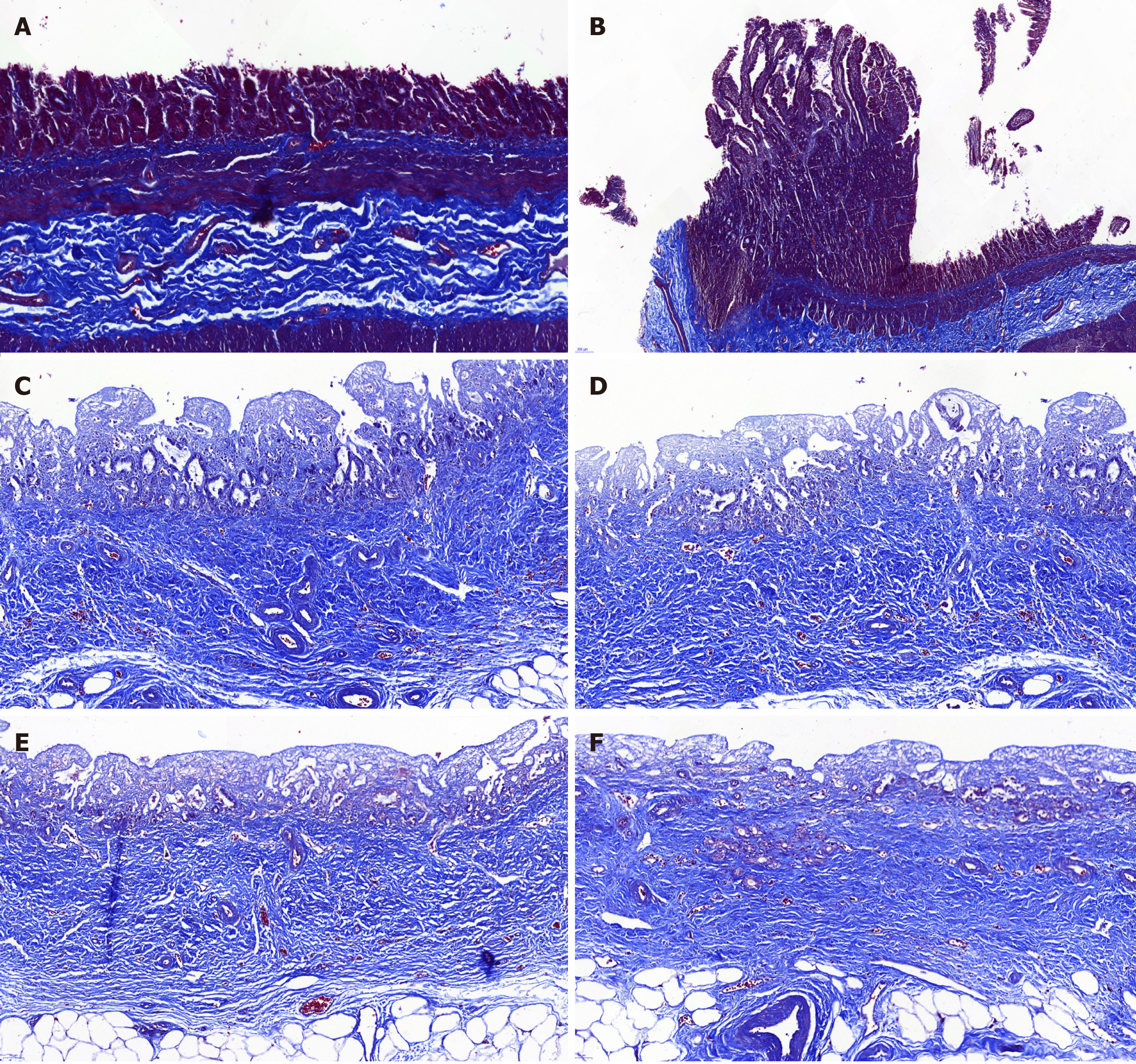
Figure 7 Masson’s trichrome staining.
The Masson’s trichrome staining showed marked regeneration of the submucosal connective tissue and no significant differences between the animal-derived artificial bile duct site and the normal bile duct. The submucosal connective tissue was more regular and thicker in group B than that in group A. A: Masson staining of the animal-derived artificial bile duct site in group A (magnification: 20 ×); B: Masson staining of duodenal papilla in group A (magnification: 20 ×); C and D: Masson staining of the animal-derived artificial bile duct site in group B (magnification: 20 ×); E and F: Masson staining of the distal normal bile duct in group B (magnification: 20 ×).
- Citation: Shang H, Zeng JP, Wang SY, Xiao Y, Yang JH, Yu SQ, Liu XC, Jiang N, Shi XL, Jin S. Extrahepatic bile duct reconstruction in pigs with heterogenous animal-derived artificial bile ducts: A preliminary experience. World J Gastroenterol 2020; 26(46): 7312-7324
- URL: https://www.wjgnet.com/1007-9327/full/v26/i46/7312.htm
- DOI: https://dx.doi.org/10.3748/wjg.v26.i46.7312


