Copyright
©The Author(s) 2020.
World J Gastroenterol. Dec 7, 2020; 26(45): 7263-7271
Published online Dec 7, 2020. doi: 10.3748/wjg.v26.i45.7263
Published online Dec 7, 2020. doi: 10.3748/wjg.v26.i45.7263
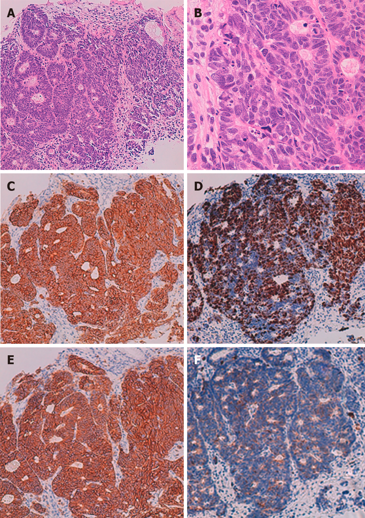
Figure 3 Histological findings of a mucosal lesion.
A: Low-power histological view of Hematoxylin and eosin (H-E) stained specimens showing atypical epithelial cells with solid alveolar nests; B: High-power view of H-E stained specimens reveals poorly differentiated atypical cells with large nucleus-cytoplasm ratio; C: Synaptophysin positive; D: Ki 67 index is approximately 70%; E: CD56 positive; F: Chromogranin A positive.
- Citation: Ishida N, Miyazu T, Tamura S, Suzuki S, Tani S, Yamade M, Iwaizumi M, Osawa S, Hamaya Y, Shinmura K, Sugimura H, Miura K, Furuta T, Sugimoto K. Tuberous sclerosis patient with neuroendocrine carcinoma of the esophagogastric junction: A case report. World J Gastroenterol 2020; 26(45): 7263-7271
- URL: https://www.wjgnet.com/1007-9327/full/v26/i45/7263.htm
- DOI: https://dx.doi.org/10.3748/wjg.v26.i45.7263


