Copyright
©The Author(s) 2020.
World J Gastroenterol. Nov 14, 2020; 26(42): 6658-6668
Published online Nov 14, 2020. doi: 10.3748/wjg.v26.i42.6658
Published online Nov 14, 2020. doi: 10.3748/wjg.v26.i42.6658
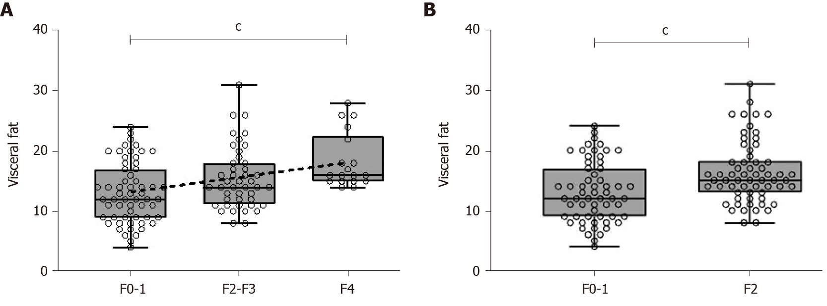
Figure 1 Visceral fat measurement by bioimpedanciometry, according to histological fibrosis stage.
A: Visceral fat measurements increased along with fibrosis stage assessed by histological analysis (F0-1, 12; F2-3, 14; F4, 16; Kruskal-Wallis cP < 0.001). A line can be fit by linear regression, showing linear association (r2 = 0.11, cP < 0.001); B: Visceral fat measurements were greater for those patients with significant fibrosis (16.3 vs 13.1, cP < 0.001).
- Citation: Hernández-Conde M, Llop E, Fernández Carrillo C, Tormo B, Abad J, Rodriguez L, Perelló C, López Gomez M, Martínez-Porras JL, Fernández Puga N, Trapero-Marugan M, Fraga E, Ferre Aracil C, Calleja Panero JL. Estimation of visceral fat is useful for the diagnosis of significant fibrosis in patients with non-alcoholic fatty liver disease. World J Gastroenterol 2020; 26(42): 6658-6668
- URL: https://www.wjgnet.com/1007-9327/full/v26/i42/6658.htm
- DOI: https://dx.doi.org/10.3748/wjg.v26.i42.6658


