Copyright
©The Author(s) 2020.
World J Gastroenterol. Nov 14, 2020; 26(42): 6614-6625
Published online Nov 14, 2020. doi: 10.3748/wjg.v26.i42.6614
Published online Nov 14, 2020. doi: 10.3748/wjg.v26.i42.6614
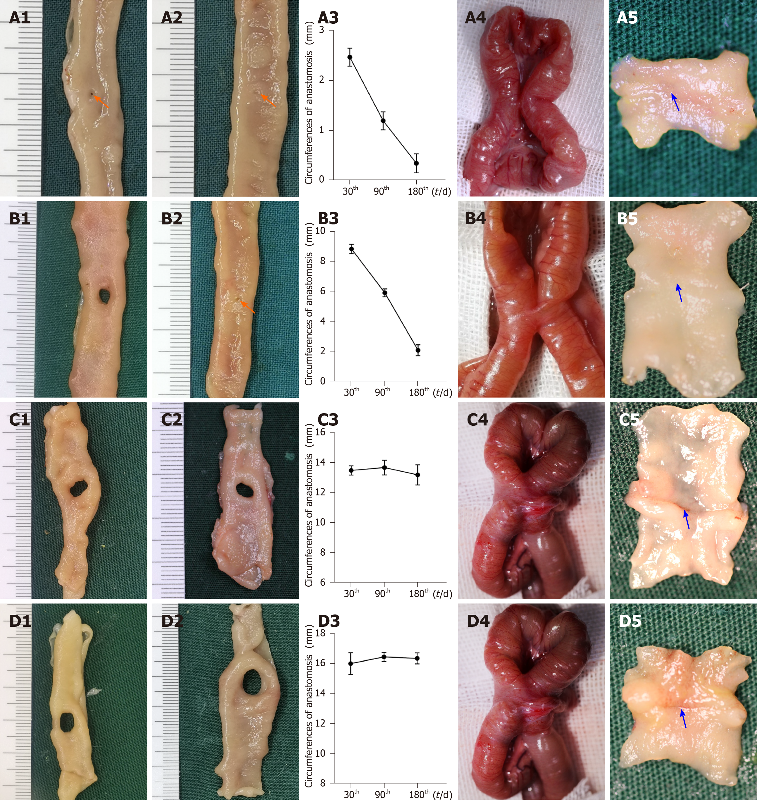
Figure 3 Gross appearance of anastomoses using traditional nummular magnetic compression anastomosis devices.
A: Group 1.1 (Φ3 mm): The size of anastomosis 30 d after magnetic compression anastomosis (MCA) (A1), the size of anastomosis 180 d after MCA (A2), the change in anastomosis circumferences after MCA (A3), serosa side of anastomosis (A4), and mucosa side of anastomosis (A5); B: Group 1.2 (Φ4 mm): The size of anastomosis 30 d after MCA (B1), the size of anastomosis 180 d after MCA (B2), the change in anastomosis circumferences after MCA (B3), serosa side of anastomosis (B4), and mucosa side of anastomosis (B5); C: Group 1.3 (Φ5 mm): The size of anastomosis 30 d after MCA (C1), the size of anastomosis 180 d after MCA (C2), the change in anastomosis circumferences after MCA (C3), serosa side of anastomosis (C4), and mucosa side of anastomosis (C5); D: Group 1.4 (Φ6 mm): The size of anastomosis 30 d after MCA (D1), the size of anastomosis 180 d after MCA (D2), the change in anastomosis circumferences after MCA (D3), serosa side of anastomosis (D4), and mucosa side of anastomosis (D5). Orange arrows: Anastomosis; blue arrows: Anastomotic line.
- Citation: Chen H, Ma T, Wang Y, Zhu HY, Feng Z, Wu RQ, Lv Y, Dong DH. Fedora-type magnetic compression anastomosis device for intestinal anastomosis. World J Gastroenterol 2020; 26(42): 6614-6625
- URL: https://www.wjgnet.com/1007-9327/full/v26/i42/6614.htm
- DOI: https://dx.doi.org/10.3748/wjg.v26.i42.6614


