Copyright
©The Author(s) 2020.
World J Gastroenterol. Nov 14, 2020; 26(42): 6582-6598
Published online Nov 14, 2020. doi: 10.3748/wjg.v26.i42.6582
Published online Nov 14, 2020. doi: 10.3748/wjg.v26.i42.6582
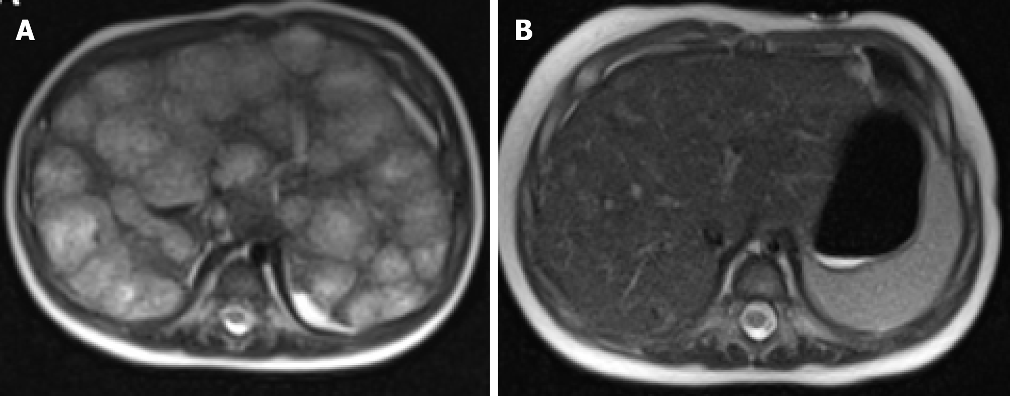
Figure 11 Magnetic resonance imaging results.
A: Magnetic resonance imaging (MRI) of liver in a 4-month-old at the time of the diagnosis, axial T2-weighted (HASTE) image showing multiple nodular hyperintense lesions with centripetal fill-in on the delayed phase; B: MRI of the liver 3 mo after starting atenolol treatment, subsequent axial T2-weighted image showing interval decrease in size of enhancing lesions and improving hepatomegaly.
- Citation: Schmalz MJ, Radhakrishnan K. Vascular anomalies associated with hepatic shunting. World J Gastroenterol 2020; 26(42): 6582-6598
- URL: https://www.wjgnet.com/1007-9327/full/v26/i42/6582.htm
- DOI: https://dx.doi.org/10.3748/wjg.v26.i42.6582


