Copyright
©The Author(s) 2020.
World J Gastroenterol. Nov 14, 2020; 26(42): 6582-6598
Published online Nov 14, 2020. doi: 10.3748/wjg.v26.i42.6582
Published online Nov 14, 2020. doi: 10.3748/wjg.v26.i42.6582
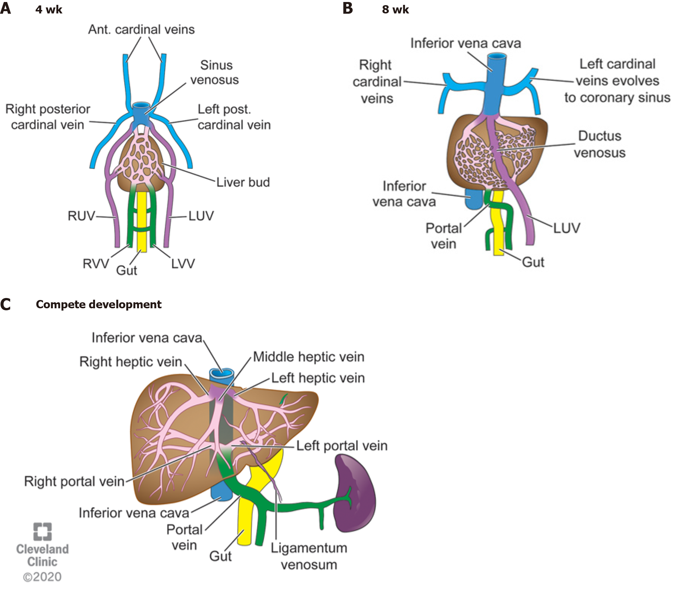
Figure 1 Four weeks gestation, eight weeks gestation, and the mature liver vasculature after birth.
A: Right and left umbilical, cardinal, and vitelline veins making up the primitive vasculature to the developing liver bud. Cardinal and umbilical veins converge on top of the liver to form the sinus venosus. The vitelline veins return blood from the developing gut; B: Posterior cardinal veins coalesce to form the upper part of the inferior vena cava. The right umbilical vein involutes and left umbilical vein makes up the ductus venosus (DV). The intrahepatic vessels start to form mature hepatic veins. The vitelline veins start to mature into the portal venous system; C: The DV collapses at birth after the umbilical cord is cut and becomes the ligamentum venosum. The portal veins and hepatic veins are mature. RUV: Right umbilical vein; LUV: Left umbilical vein; RRV: Right vitelline vein; LVV: Left vitelline vein; IVC: Inferior vena cava. Reprinted with permission, Cleveland Clinic Center for Medical Art & Photography ©2020. All rights reserved.
- Citation: Schmalz MJ, Radhakrishnan K. Vascular anomalies associated with hepatic shunting. World J Gastroenterol 2020; 26(42): 6582-6598
- URL: https://www.wjgnet.com/1007-9327/full/v26/i42/6582.htm
- DOI: https://dx.doi.org/10.3748/wjg.v26.i42.6582


