Copyright
©The Author(s) 2020.
World J Gastroenterol. Nov 7, 2020; 26(41): 6431-6441
Published online Nov 7, 2020. doi: 10.3748/wjg.v26.i41.6431
Published online Nov 7, 2020. doi: 10.3748/wjg.v26.i41.6431
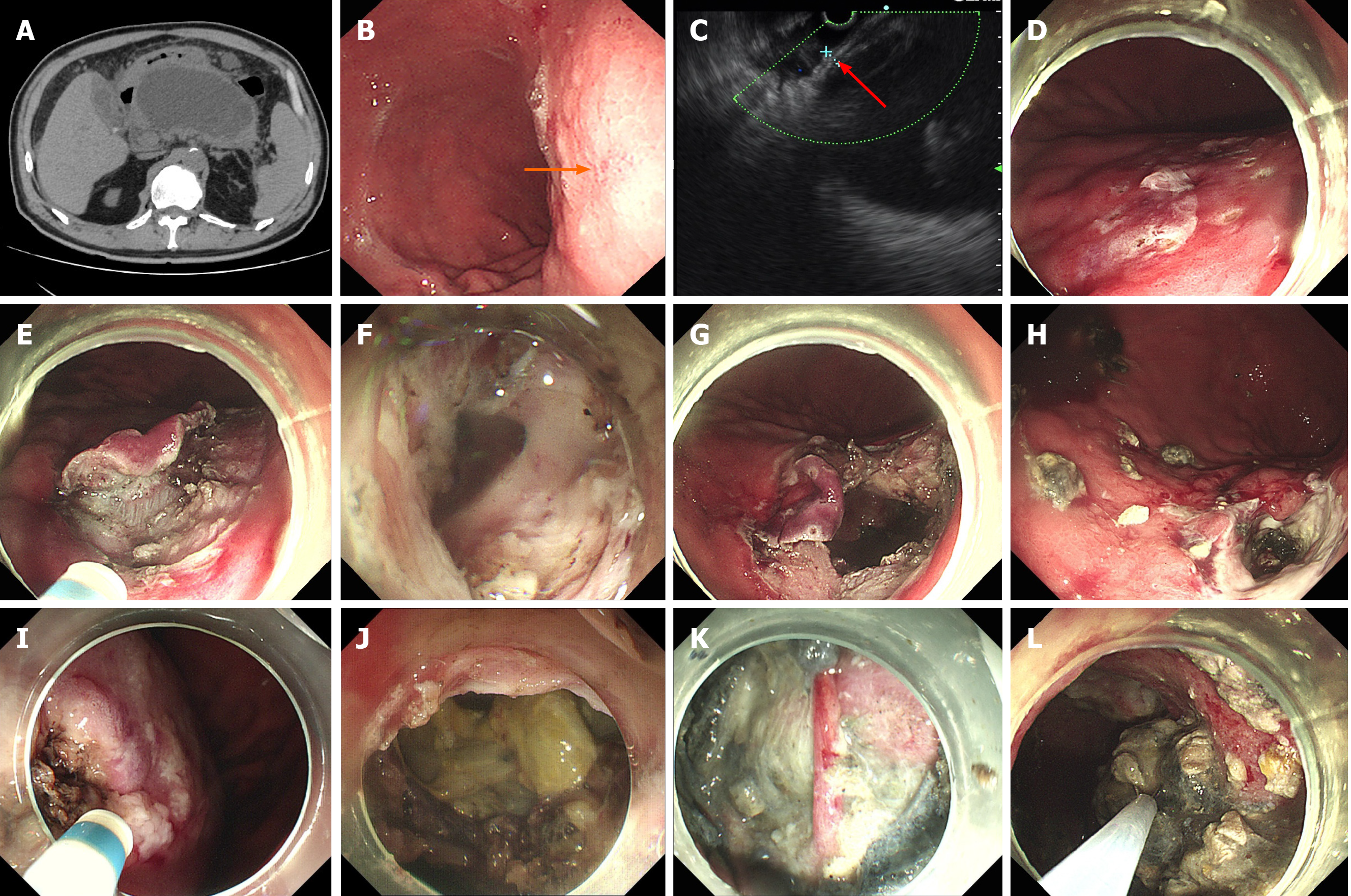
Figure 2 Endoscopic gastric fenestration technique.
A: Closely connected walled-off necrosis (WON) and gastric wall lacking clear layers (black arrow, preoperative computed tomography scan); B: Compressive indentation of stomach by WON, with intense inflammation (orange arrow); C: Endoscopic ultrasound assessment and selection of fenestration site, abutment < 1 cm in combined thickness without clear layers (red arrow); D: Marking of prospective fenestration; E: Initial fenestration by endoscopic submucosal dissection; F: Penetration of WON capsule, releasing fluid content; G: Expanded fenestration; H: Self-healing of fenestration as seen by postoperative endoscopy (1 wk after endoscopic gastric fenestration); I: Narrowed area of initial fenestration; J: Enlarged expanded fenestration up to 3 cm; K: Necrotic tissue and exposed blood vessel in WON; L: Debridement of necrotic tissue.
- Citation: Liu F, Wu L, Wang XD, Xiao JG, Li W. Endoscopic gastric fenestration of debriding pancreatic walled-off necrosis: A pilot study. World J Gastroenterol 2020; 26(41): 6431-6441
- URL: https://www.wjgnet.com/1007-9327/full/v26/i41/6431.htm
- DOI: https://dx.doi.org/10.3748/wjg.v26.i41.6431


