Copyright
©The Author(s) 2020.
World J Gastroenterol. Oct 28, 2020; 26(40): 6163-6181
Published online Oct 28, 2020. doi: 10.3748/wjg.v26.i40.6163
Published online Oct 28, 2020. doi: 10.3748/wjg.v26.i40.6163
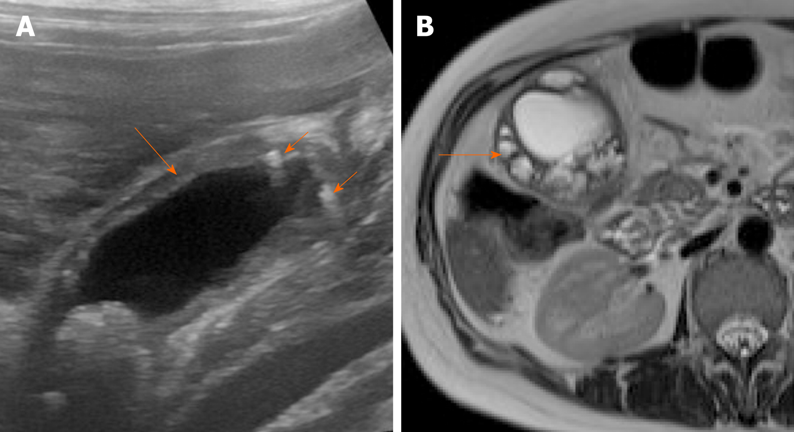
Figure 7 Imaging findings in adenomyomatosis.
A: Ultrasound image shows symmetrical mural thickening (arrow) with echogenic foci showing comet tail artifacts (short arrows); B: T2-weighted magnetic resonance imaging shows multiple intramural cystic lesions (arrow).
- Citation: Gupta P, Marodia Y, Bansal A, Kalra N, Kumar-M P, Sharma V, Dutta U, Sandhu MS. Imaging-based algorithmic approach to gallbladder wall thickening. World J Gastroenterol 2020; 26(40): 6163-6181
- URL: https://www.wjgnet.com/1007-9327/full/v26/i40/6163.htm
- DOI: https://dx.doi.org/10.3748/wjg.v26.i40.6163


