Copyright
©The Author(s) 2020.
World J Gastroenterol. Oct 28, 2020; 26(40): 6163-6181
Published online Oct 28, 2020. doi: 10.3748/wjg.v26.i40.6163
Published online Oct 28, 2020. doi: 10.3748/wjg.v26.i40.6163
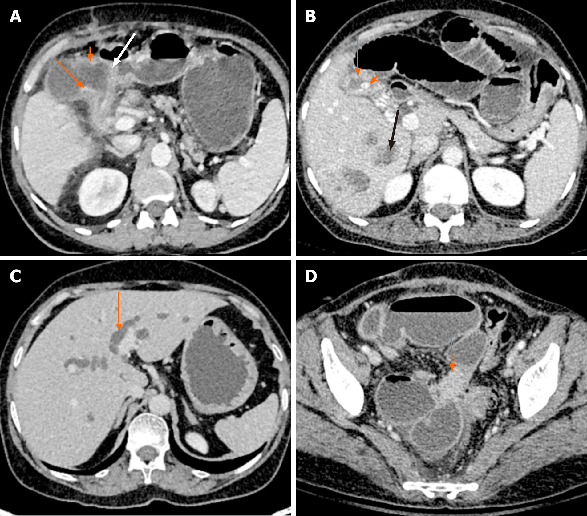
Figure 3 Direct and indirect findings of malignant gallbladder wall thickening on computed tomography.
A: There is asymmetrical mural thickening of the gallbladder neck (arrow). Also note the nodularity of the gallbladder wall in the body (short arrow) and loss of fat plane with the antropyloric region of stomach (white arrow); B: Gallbladder wall is thickened and heterogeneous (arrow). There is a calculus at the neck (short arrow). Multiple hypodense lesions are seen in right lobe (black arrow). Fine needle aspiration cytology from one of these lesions revealed metastasis; C: There is bilobar intrahepatic biliary radicle dilatation in a patient with gallbladder cancer (arrow); D: A soft tissue deposit is seen in subserosal location in pelvis causing bowel obstruction (arrow).
- Citation: Gupta P, Marodia Y, Bansal A, Kalra N, Kumar-M P, Sharma V, Dutta U, Sandhu MS. Imaging-based algorithmic approach to gallbladder wall thickening. World J Gastroenterol 2020; 26(40): 6163-6181
- URL: https://www.wjgnet.com/1007-9327/full/v26/i40/6163.htm
- DOI: https://dx.doi.org/10.3748/wjg.v26.i40.6163


