Copyright
©The Author(s) 2020.
World J Gastroenterol. Jan 28, 2020; 26(4): 383-392
Published online Jan 28, 2020. doi: 10.3748/wjg.v26.i4.383
Published online Jan 28, 2020. doi: 10.3748/wjg.v26.i4.383
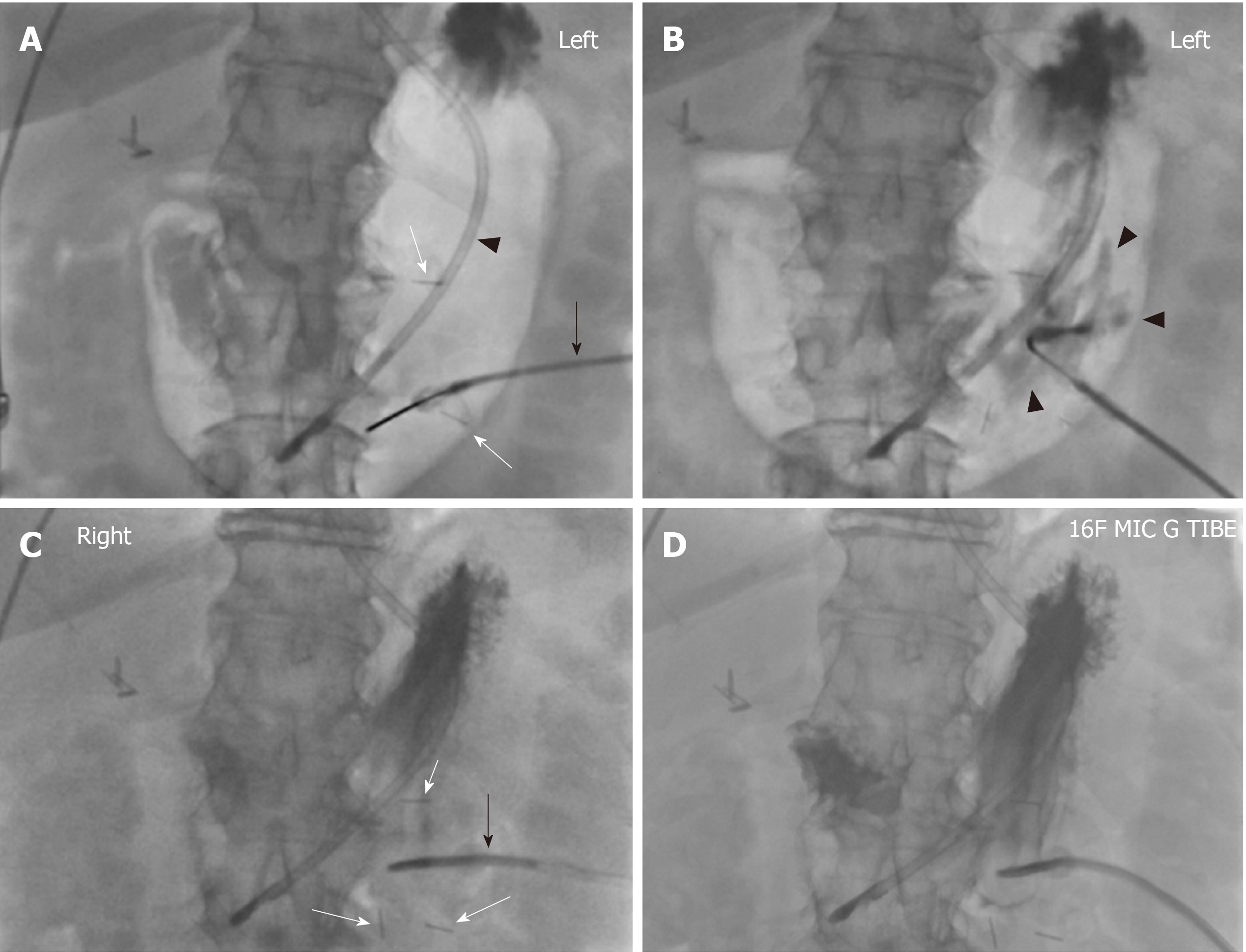
Figure 3 Seventy-six-year-old female with a history of upper extremity edema, intracranial hemorrhage and dysphagia referred for placement of percutaneous gastrostomy tube due to dysphagia with high aspiration risk.
A 16-French gastrostomy tube was placed through a 20-French peel-away sheath. The procedure time was 23 min. Fluoroscopy time was 3 min 24 s and Air Kerma was 43 mGy. No challenge was encountered during the procedure and the gastrostomy tube was started to be used for enteral feeding 24 h after the procedure. A: Dobhoff tube (arrowhead) with its tip in the gastric body was placed before the patient came to the procedure suite. The stomach was manually inflated with air. T-bar fasteners (white arrow) were advanced into the gastric body. Note the connected tubing (black arrow) which contained iodinated contrast; B: Iodinated contrast was injected into the gastric body through the T-bar fastener to confirm intraluminal location. Note the outlines of gastric rugae (black arrowheads); C: The three T-bar fasteners (white arrows) are positioned in a triangular fashion. Note the gastrostomy tube (black arrow) was angulated towards the antrum and pylorus facilitating later conversion to a gastrojejunostomy tube; D: Contrast injection through the gastrostomy tube ascertains the intragastric placement of the tube.
- Citation: Partovi S, Li X, Moon E, Thompson D. Image guided percutaneous gastrostomy catheter placement: How we do it safely and efficiently. World J Gastroenterol 2020; 26(4): 383-392
- URL: https://www.wjgnet.com/1007-9327/full/v26/i4/383.htm
- DOI: https://dx.doi.org/10.3748/wjg.v26.i4.383


