Copyright
©The Author(s) 2020.
World J Gastroenterol. Oct 14, 2020; 26(38): 5884-5895
Published online Oct 14, 2020. doi: 10.3748/wjg.v26.i38.5884
Published online Oct 14, 2020. doi: 10.3748/wjg.v26.i38.5884
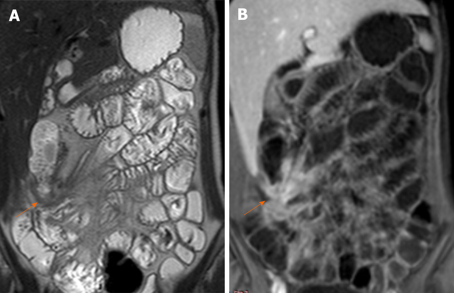
Figure 2 Magnetic resonance enterography.
A: Coronal T2WI shows enteroenteric fistula at the right iliac fossa with stellate appearance of the thickened ileal loops (white arrow); B: Coronal fat-suppressed three-dimensional gradient echo postcontrast T1WI shows accentuated star-like enhancement at the right iliac fossa denoting fistulizing crohn’s disease.
- Citation: Kamel S, Sakr M, Hamed W, Eltabbakh M, Askar S, Bassuny A, Hussein R, Elbaz A. Comparative study between bowel ultrasound and magnetic resonance enterography among Egyptian inflammatory bowel disease patients. World J Gastroenterol 2020; 26(38): 5884-5895
- URL: https://www.wjgnet.com/1007-9327/full/v26/i38/5884.htm
- DOI: https://dx.doi.org/10.3748/wjg.v26.i38.5884


