Copyright
©The Author(s) 2020.
World J Gastroenterol. Oct 14, 2020; 26(38): 5884-5895
Published online Oct 14, 2020. doi: 10.3748/wjg.v26.i38.5884
Published online Oct 14, 2020. doi: 10.3748/wjg.v26.i38.5884
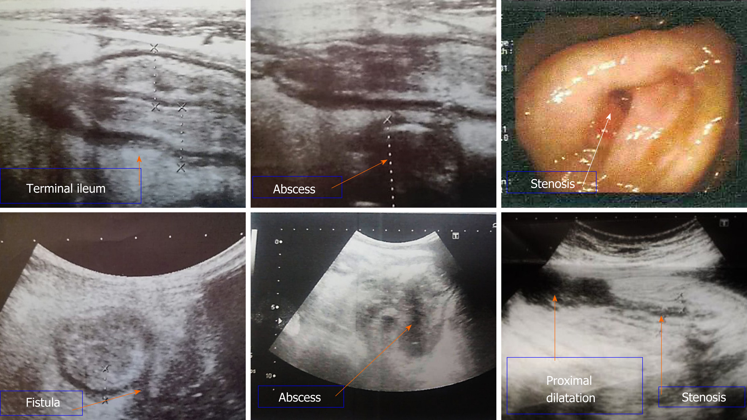
Figure 1 Bowel ultrasound and colonoscopy images.
Bowel ultrasound demonstrates diffuse terminal ileal wall thickening likely of inflammatory nature with sonographic evidence of fistulization with mesenteric abscess formation. Stenosis which was detected during colonoscopy was seen by bowel ultrasound with proximal dilatation.
- Citation: Kamel S, Sakr M, Hamed W, Eltabbakh M, Askar S, Bassuny A, Hussein R, Elbaz A. Comparative study between bowel ultrasound and magnetic resonance enterography among Egyptian inflammatory bowel disease patients. World J Gastroenterol 2020; 26(38): 5884-5895
- URL: https://www.wjgnet.com/1007-9327/full/v26/i38/5884.htm
- DOI: https://dx.doi.org/10.3748/wjg.v26.i38.5884


