Copyright
©The Author(s) 2020.
World J Gastroenterol. Oct 14, 2020; 26(38): 5784-5796
Published online Oct 14, 2020. doi: 10.3748/wjg.v26.i38.5784
Published online Oct 14, 2020. doi: 10.3748/wjg.v26.i38.5784
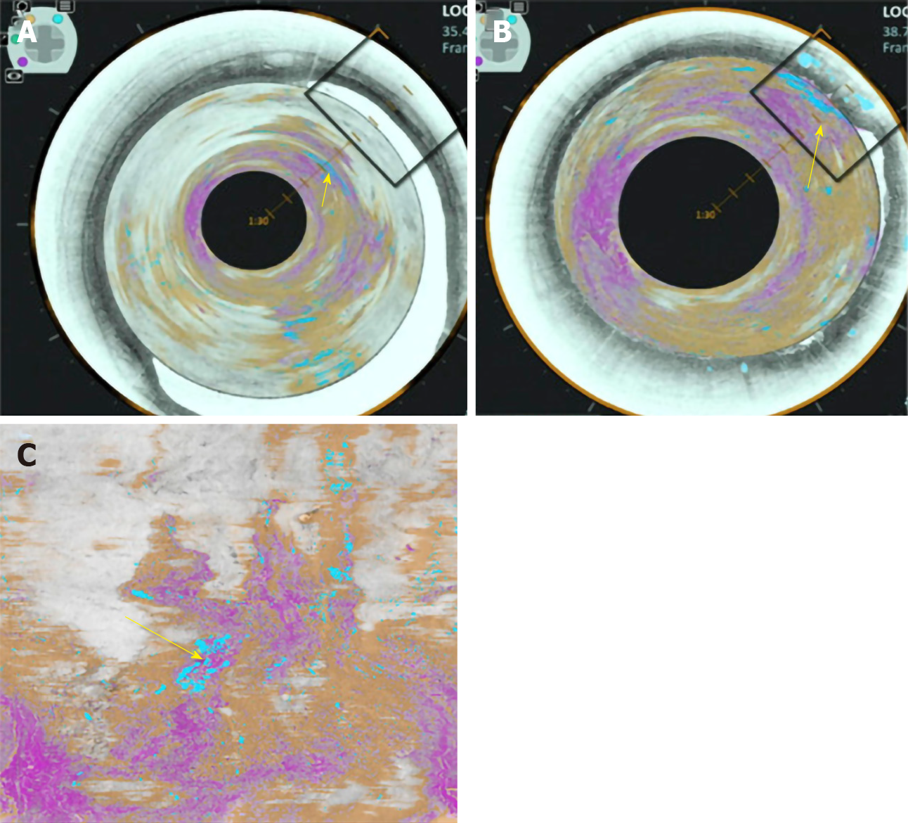
Figure 5 Volumetric laser endomicroscopy image showing area of overlap (yellow arrow) between the 3 features of dysplasia identified with the colour schemes.
A: View looking down into the oesophagus; B: Close up of dysplastic area; C: Forward view of the dysplastic area. A-C: Citation: Trindade AJ, McKinley MJ, Fan C, Leggett CL, Kahn A, Pleskow DK. Endoscopic Surveillance of Barrett's Esophagus Using Volumetric Laser Endomicroscopy With Artificial Intelligence Image Enhancement. Gastroenterology 2019; 157: 303-305. Copyright© The Authors 2019. Published by Elsevier.
- Citation: Hussein M, González-Bueno Puyal J, Mountney P, Lovat LB, Haidry R. Role of artificial intelligence in the diagnosis of oesophageal neoplasia: 2020 an endoscopic odyssey. World J Gastroenterol 2020; 26(38): 5784-5796
- URL: https://www.wjgnet.com/1007-9327/full/v26/i38/5784.htm
- DOI: https://dx.doi.org/10.3748/wjg.v26.i38.5784


