Copyright
©The Author(s) 2020.
World J Gastroenterol. Oct 7, 2020; 26(37): 5597-5605
Published online Oct 7, 2020. doi: 10.3748/wjg.v26.i37.5597
Published online Oct 7, 2020. doi: 10.3748/wjg.v26.i37.5597
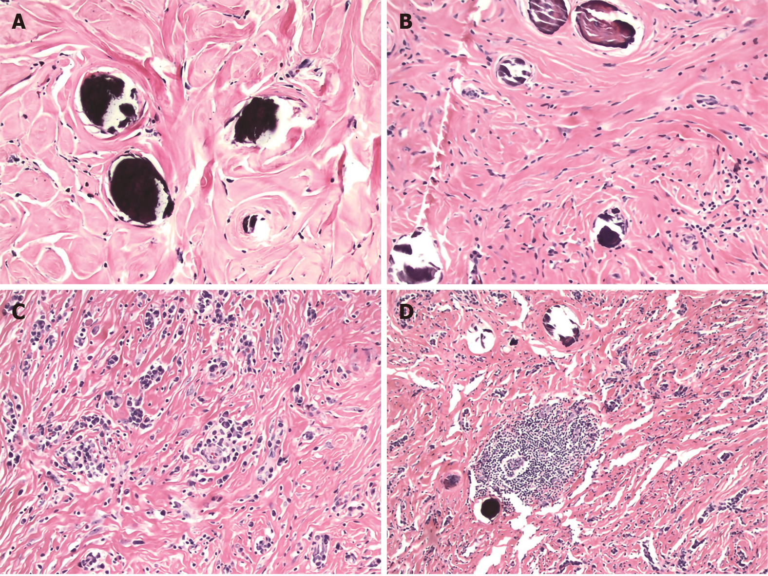
Figure 2 Histologic features of calcifying fibrous tumor.
A: Haphazard or whorled pattern hyalinization admixed with calcifications (original magnification 200 ×); B: Dense hyalinization admixed with spindle cells and psammomatous calcification (original magnification 200 ×); C: Lymphoplasmacytic infiltrate in a background of dense hyalinization (original magnification 200 ×); D: Lymphoid follicle in a background of dense hyalinization and scattered calcification (original magnification 100 ×). Hematoxylin-eosin stain.
- Citation: Turbiville D, Zhang X. Calcifying fibrous tumor of the gastrointestinal tract: A clinicopathologic review and update. World J Gastroenterol 2020; 26(37): 5597-5605
- URL: https://www.wjgnet.com/1007-9327/full/v26/i37/5597.htm
- DOI: https://dx.doi.org/10.3748/wjg.v26.i37.5597


