Copyright
©The Author(s) 2020.
World J Gastroenterol. Sep 28, 2020; 26(36): 5527-5533
Published online Sep 28, 2020. doi: 10.3748/wjg.v26.i36.5527
Published online Sep 28, 2020. doi: 10.3748/wjg.v26.i36.5527
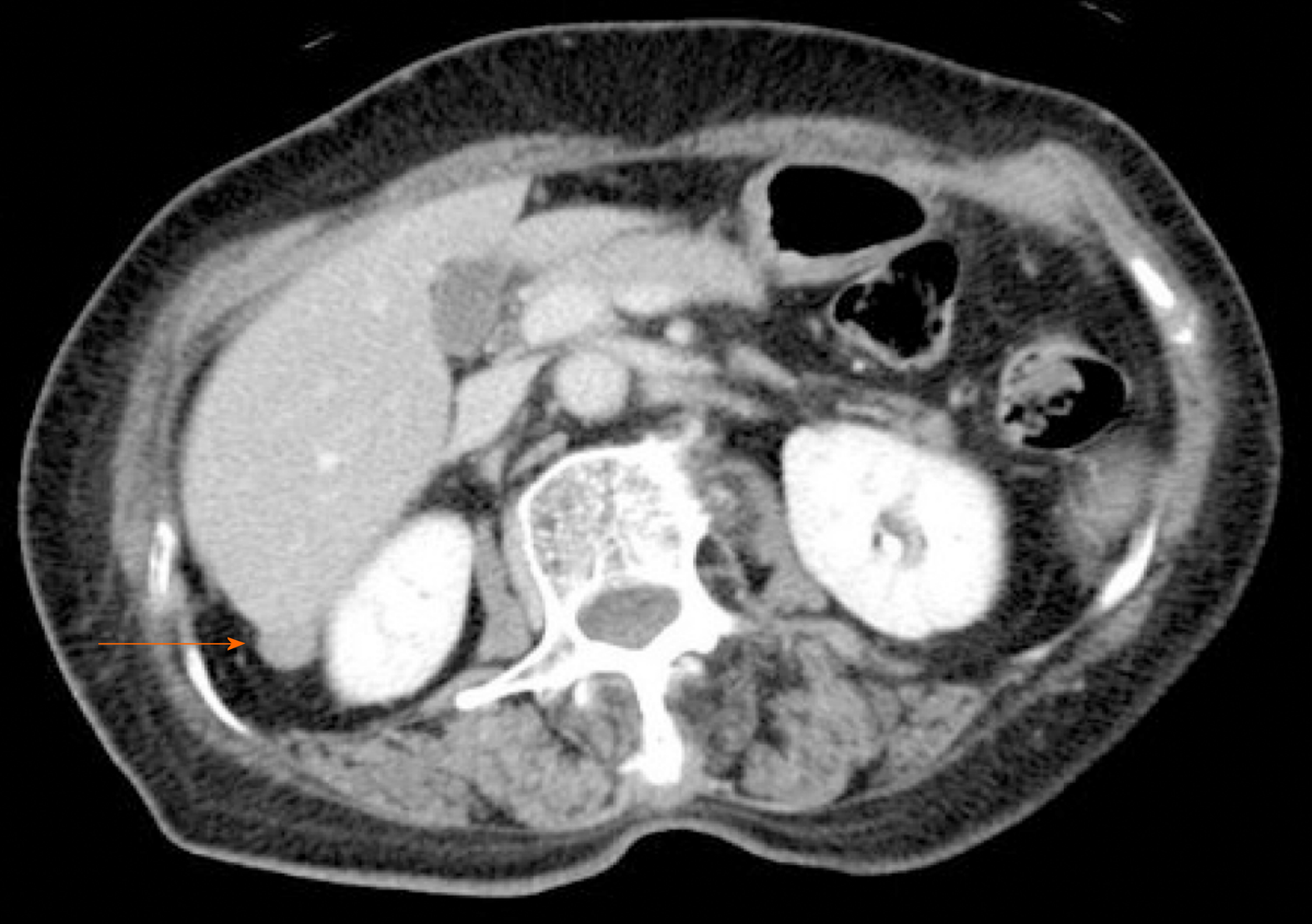
Figure 1 Contrast-enhanced computed tomography.
Contrast-enhanced computed tomography showing a slightly enhanced mass (diameter: 12 mm) located between the dorsal side of the right hepatic lobe and right kidney.
- Citation: Sugiyama Y, Shimbara K, Sasaki M, Kouyama M, Tazaki T, Takahashi S, Nakamitsu A. Solitary peritoneal metastasis of gastrointestinal stromal tumor: A case report. World J Gastroenterol 2020; 26(36): 5527-5533
- URL: https://www.wjgnet.com/1007-9327/full/v26/i36/5527.htm
- DOI: https://dx.doi.org/10.3748/wjg.v26.i36.5527


