Copyright
©The Author(s) 2020.
World J Gastroenterol. Sep 28, 2020; 26(36): 5450-5462
Published online Sep 28, 2020. doi: 10.3748/wjg.v26.i36.5450
Published online Sep 28, 2020. doi: 10.3748/wjg.v26.i36.5450
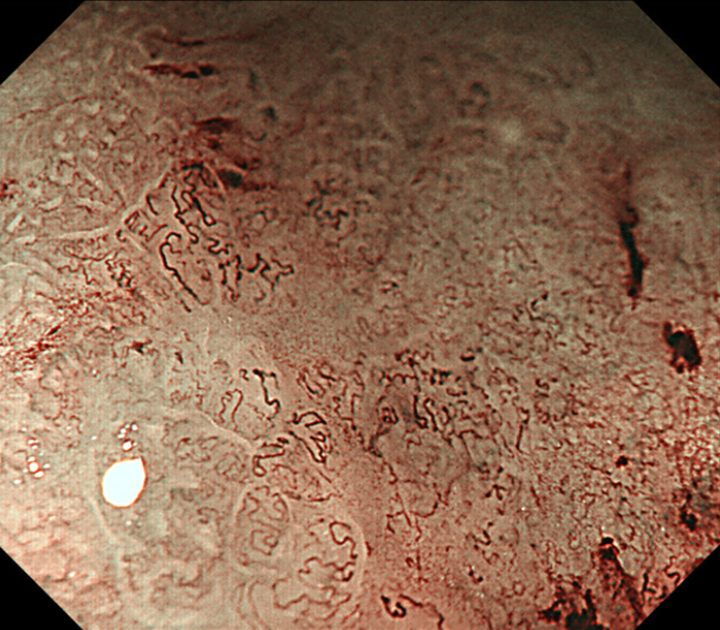
Figure 4 A representative magnifying endoscopy with narrow-band imaging image of the undifferentiated-type component.
Magnifying endoscopy with narrow-band imaging finding of undifferentiated-type. Component was defined as satisfying both a microvascular pattern, which has an isolated and disordered quality and a microstructure pattern, which lacks visibility or even disappears. It complied with the finding described as a corkscrew pattern.
- Citation: Ozeki Y, Hirasawa K, Kobayashi R, Sato C, Tateishi Y, Sawada A, Ikeda R, Nishio M, Fukuchi T, Makazu M, Taguri M, Maeda S. Histopathological validation of magnifying endoscopy for diagnosis of mixed-histological-type early gastric cancer. World J Gastroenterol 2020; 26(36): 5450-5462
- URL: https://www.wjgnet.com/1007-9327/full/v26/i36/5450.htm
- DOI: https://dx.doi.org/10.3748/wjg.v26.i36.5450


