Copyright
©The Author(s) 2020.
World J Gastroenterol. Sep 28, 2020; 26(36): 5450-5462
Published online Sep 28, 2020. doi: 10.3748/wjg.v26.i36.5450
Published online Sep 28, 2020. doi: 10.3748/wjg.v26.i36.5450
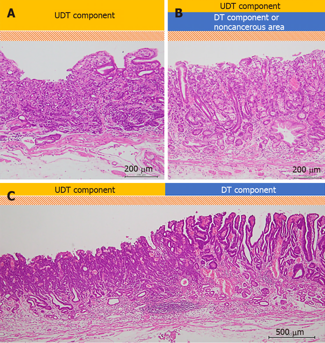
Figure 2 Evaluation of histopathological examination (hematoxylin-eosin staining).
A schema of the three histopathological findings investigated in our study. The scale bars show 200 μm in (A) and (B), and 500 μm in (C). A: The whole mucosal layer occupation of undifferentiated-type (UDT) component (100 ×); B: Exposure of UDT component to the mucosal surface (100 ×); C: Clear border between differentiated-type and UDT components (40 ×). DT: Differentiated-type; UDT: Undifferentiated-type.
- Citation: Ozeki Y, Hirasawa K, Kobayashi R, Sato C, Tateishi Y, Sawada A, Ikeda R, Nishio M, Fukuchi T, Makazu M, Taguri M, Maeda S. Histopathological validation of magnifying endoscopy for diagnosis of mixed-histological-type early gastric cancer. World J Gastroenterol 2020; 26(36): 5450-5462
- URL: https://www.wjgnet.com/1007-9327/full/v26/i36/5450.htm
- DOI: https://dx.doi.org/10.3748/wjg.v26.i36.5450


