Copyright
©The Author(s) 2020.
World J Gastroenterol. Sep 7, 2020; 26(33): 4945-4959
Published online Sep 7, 2020. doi: 10.3748/wjg.v26.i33.4945
Published online Sep 7, 2020. doi: 10.3748/wjg.v26.i33.4945
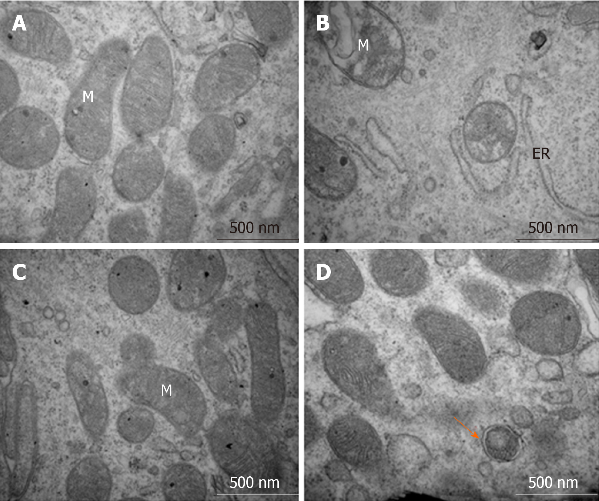
Figure 7 Structures of the intestinal epithelial cells and autolysosomes observed by transmission electron microscopy.
A: Control group showed normal organelle structure; B: Dextran sulfate sodium induced colitis group showed mitochondrial swelling and ruptures of internal cristae, along with distention of the rough endoplasmic reticulum; C: 5-aminosalicylic acid group revealed that mitochondrial swelling was not obvious, and mitochondrial cristae showed blurred appearance; and D: Resveratrol treatment group showed that organelle structure was basically normal, and autophagosome could be observed. ER: Endoplasmic reticulum, M: Mitochondria.
- Citation: Pan HH, Zhou XX, Ma YY, Pan WS, Zhao F, Yu MS, Liu JQ. Resveratrol alleviates intestinal mucosal barrier dysfunction in dextran sulfate sodium-induced colitis mice by enhancing autophagy. World J Gastroenterol 2020; 26(33): 4945-4959
- URL: https://www.wjgnet.com/1007-9327/full/v26/i33/4945.htm
- DOI: https://dx.doi.org/10.3748/wjg.v26.i33.4945


