Copyright
©The Author(s) 2020.
World J Gastroenterol. Jan 21, 2020; 26(3): 366-374
Published online Jan 21, 2020. doi: 10.3748/wjg.v26.i3.366
Published online Jan 21, 2020. doi: 10.3748/wjg.v26.i3.366
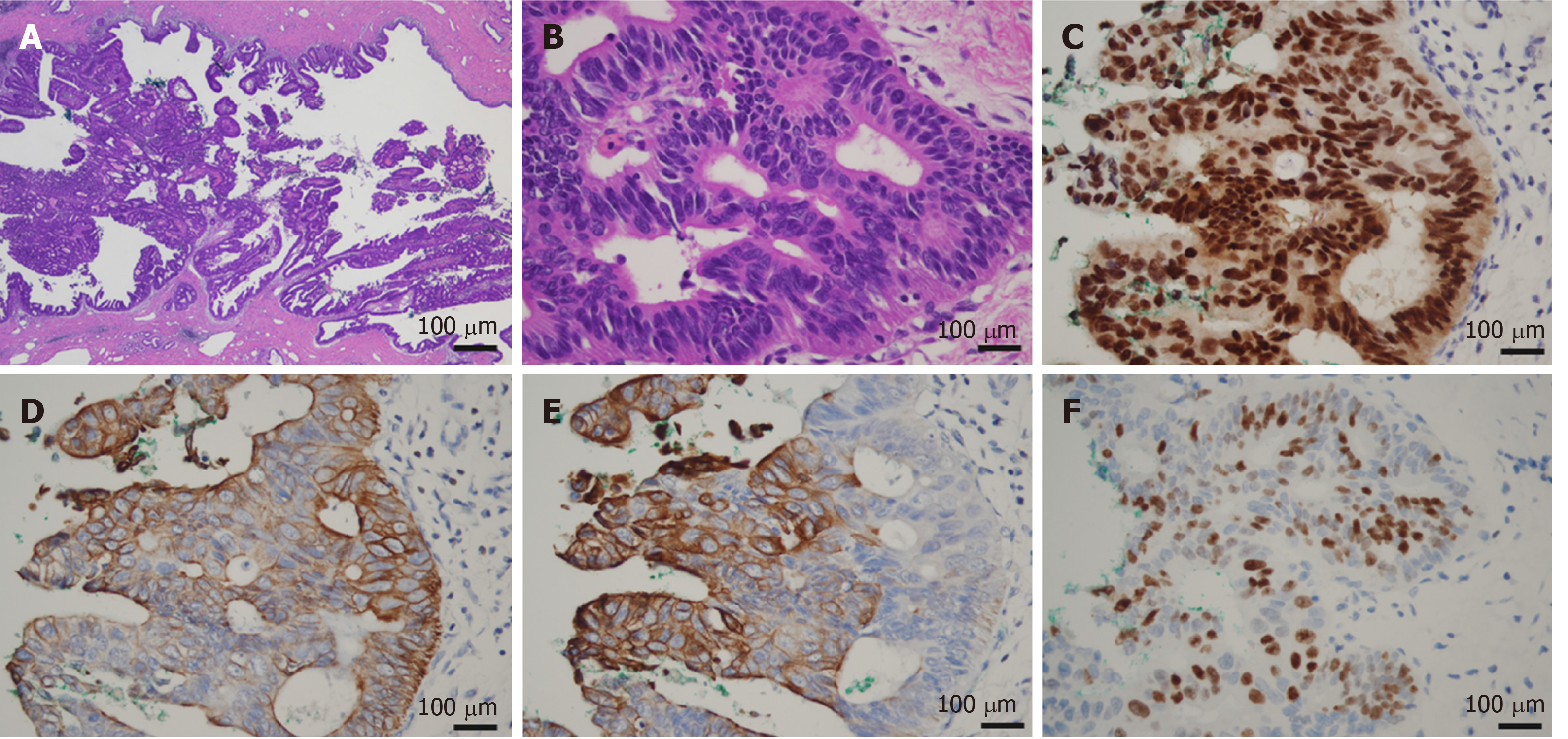
Figure 4 Microscopic findings of the bile duct tumor.
A: The tumor showed the intraductal papillary proliferation of columnar cells with a high nuclear/cytoplasmic ratio; B: These neoplastic cells were entirely confined within the duct, and no invasion was identified (A: magnification × 20, B: magnification × 400); C-F: Immunohistological staining showed that the tumor cells were positive for CDX2, CK7, CK20, and Ki67.
- Citation: Nam NH, Taura K, Kanai M, Fukuyama K, Uza N, Maeda H, Yutaka Y, Chen-Yoshikawa TF, Muto M, Uemoto S. Unexpected metastasis of intraductal papillary neoplasm of the bile duct without an invasive component to the brain and lungs: A case report. World J Gastroenterol 2020; 26(3): 366-374
- URL: https://www.wjgnet.com/1007-9327/full/v26/i3/366.htm
- DOI: https://dx.doi.org/10.3748/wjg.v26.i3.366


