Copyright
©The Author(s) 2020.
World J Gastroenterol. Jan 21, 2020; 26(3): 291-306
Published online Jan 21, 2020. doi: 10.3748/wjg.v26.i3.291
Published online Jan 21, 2020. doi: 10.3748/wjg.v26.i3.291
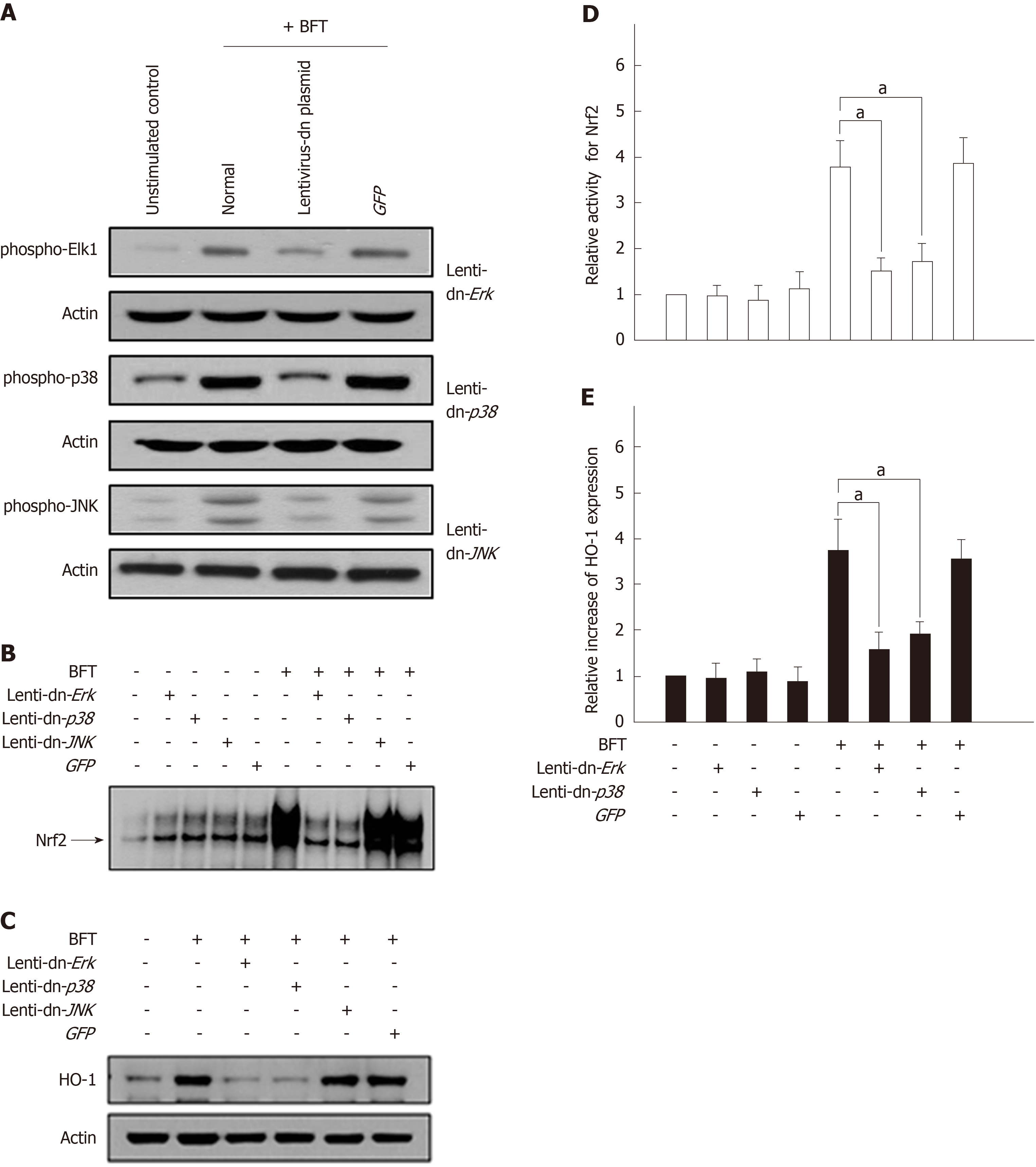
Figure 8 Effects of mitogen-activated protein kinases suppression on heme oxygenase-1 expression in dendritic cells stimulated with Bacteroides fragilis enterotoxin.
A: DC2.4 cells were infected with lentiviruses containing either a dominant-negative or control plasmid (GFP). Transfected cells were stimulated with BFT (100 ng/mL) for 30 min and immunoblots were then performed. Results are representative of three independent experiments. B and C: Transfected cells were stimulated with BFT (100 ng/mL) for 3 h (Nrf2) or 24 h (heme oxygenase-1, HO-1). B: DNA binding activities of Nrf2 were evaluated by EMSA. C: Expression of HO-1 and actin was analyzed by immunoblot. Results are representative of more than three independent experiments. D: Transfected cells were stimulated with BFT (100 ng/mL) for 3 h. Phospho-Nrf2 activities were measured using an ELISA kit. Data are expressed as mean fold induction ± SE of Nrf2 relative to untreated controls (n = 5). E: Transfected cells were stimulated with BFT (100 ng/mL) for 24 h. Transfected cells were either left untreated or stimulated with BFT (100 ng/mL) for another 6 h (Nrf2) or 24 h (HO-1). Each ELISA kit measured activities of phospho-IκBα and Nrf2, as well as HO-1 expression. Data are expressed as mean fold induction ± SE (%) relative to untreated controls (n = 5). aP < 0.05. HO-1: Heme oxygenase-1; BFT: Bacteroides fragilis toxin.
- Citation: Ko SH, Jeon JI, Woo HA, Kim JM. Bacteroides fragilis enterotoxin upregulates heme oxygenase-1 in dendritic cells via reactive oxygen species-, mitogen-activated protein kinase-, and Nrf2-dependent pathway. World J Gastroenterol 2020; 26(3): 291-306
- URL: https://www.wjgnet.com/1007-9327/full/v26/i3/291.htm
- DOI: https://dx.doi.org/10.3748/wjg.v26.i3.291


