Copyright
©The Author(s) 2020.
World J Gastroenterol. Jan 21, 2020; 26(3): 291-306
Published online Jan 21, 2020. doi: 10.3748/wjg.v26.i3.291
Published online Jan 21, 2020. doi: 10.3748/wjg.v26.i3.291
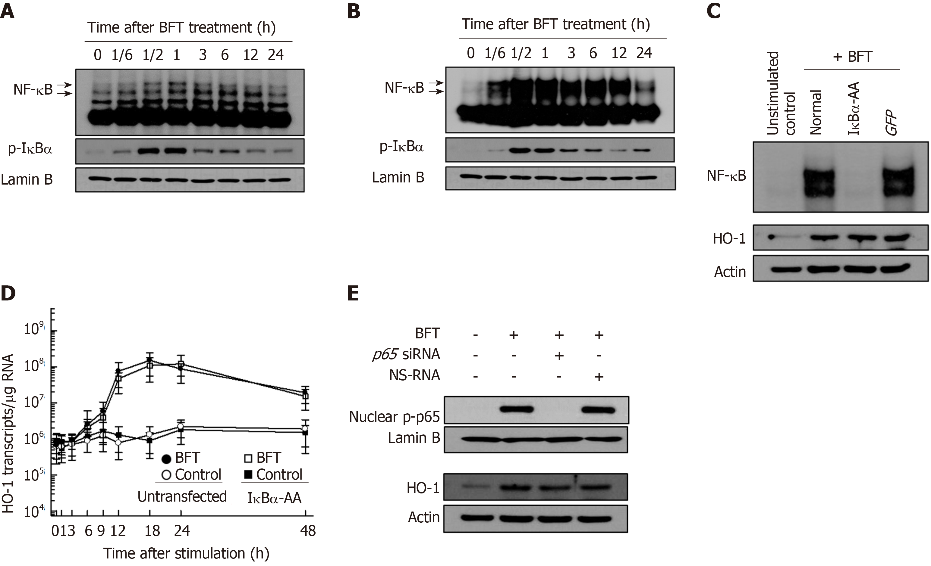
Figure 2 Effects of NF-κB suppression on heme oxygenase-1 expression in dendritic cells treated with Bacteroides fragilis enterotoxin.
A and B: Bone marrow-derived dendritic cells (DCs) (A) and DC2.4 cells (B) were treated with BFT (100 ng/mL) for the indicated times. NF-κB DNA binding activity was assessed by EMSA. Immunoblot results for concurrent phospho-IκBα and lamin B in nuclear extracts under the same conditions are provided beneath the EMSA. C: DC2.4 cells were transfected with either lentivirus containing IκBα-superrepressor (IκBα-AA) or control virus (GFP). Transfected cells were stimulated with BFT (100 ng/ml) for 1 h. NF-κB binding activity was assayed by EMSA (top panel). Transfected or untransfected cells were treated with BFT (100 μg/ml) for 12 h. Expression of heme oxygenase-1 (HO-1) and actin was analyzed by immunoblot (bottom panel). Results are representative of more than three independent experiments. D: Transfected DC2.4 cells were treated with BFT (100 ng/ml) for the indicated periods. The levels of HO-1 mRNA were analyzed by quantitative RT-PCR using a standard RNA. The values are expressed as mean ± SD (n = 5). The β-actin mRNA levels in each group remained relatively constant throughout the same periods (approximately 106 transcripts/μg total RNA). aP < 0.05 vs untransfected cells treated with BFT. E: DC2.4 cells were transfected with NF-κB p65-specific silencing siRNA or NS-RNA as a control for 48 h, after which cells were combined with BFT (100 ng/mL) for 1 h. Nuclear extracts were analyzed by immunoblotting with the indicated Abs (top panel). Transfected cells were stimulated with BFT (100 ng/mL) for 24 h. Expression of HO-1 and actin was analyzed by immunoblot (bottom panel). Results shown are representative of more than three independent experiments. HO-1: Heme oxygenase-1; BFT: Bacteroides fragilis toxin.
- Citation: Ko SH, Jeon JI, Woo HA, Kim JM. Bacteroides fragilis enterotoxin upregulates heme oxygenase-1 in dendritic cells via reactive oxygen species-, mitogen-activated protein kinase-, and Nrf2-dependent pathway. World J Gastroenterol 2020; 26(3): 291-306
- URL: https://www.wjgnet.com/1007-9327/full/v26/i3/291.htm
- DOI: https://dx.doi.org/10.3748/wjg.v26.i3.291


