Copyright
©The Author(s) 2020.
World J Gastroenterol. Aug 7, 2020; 26(29): 4372-4377
Published online Aug 7, 2020. doi: 10.3748/wjg.v26.i29.4372
Published online Aug 7, 2020. doi: 10.3748/wjg.v26.i29.4372
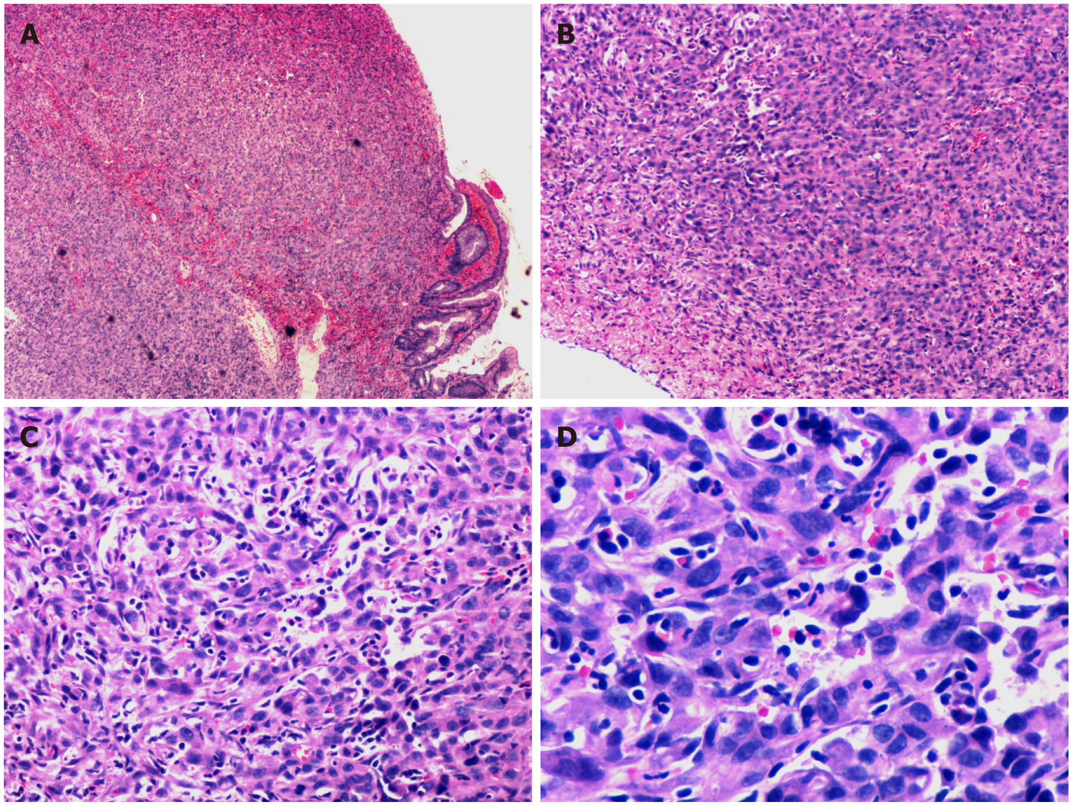
Figure 2 Histopathological findings.
Poorly differentiated neoplasms consisting of large cells with pleomorphic nuclei and abundant eosinophilic cytoplasm were observed. A: Haematoxylin and eosin (HE) staining section × 40; B: HE staining section (× 100); C: HE staining section (× 200); and D: HE staining section (× 400).
- Citation: Chen YW, Dong J, Chen WY, Dai YN. Multifocal gastrointestinal epithelioid angiosarcomas diagnosed by endoscopic mucosal resection: A case report. World J Gastroenterol 2020; 26(29): 4372-4377
- URL: https://www.wjgnet.com/1007-9327/full/v26/i29/4372.htm
- DOI: https://dx.doi.org/10.3748/wjg.v26.i29.4372


