Copyright
©The Author(s) 2020.
World J Gastroenterol. Aug 7, 2020; 26(29): 4327-4342
Published online Aug 7, 2020. doi: 10.3748/wjg.v26.i29.4327
Published online Aug 7, 2020. doi: 10.3748/wjg.v26.i29.4327
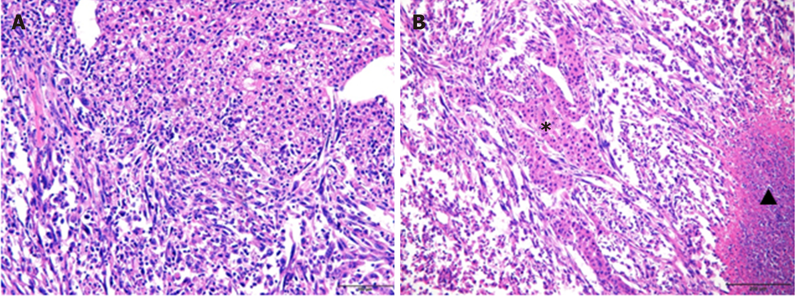
Figure 6 Pathological findings of sarcomatoid hepatocellular carcinoma.
A: The lower left area of the image shows the sarcomatous change, with spindle-shaped cells forming interlacing bundles. The upper right region represents conventional hepatocellular carcinoma, with tumor cells at Edmondson-Steiner (ES) grade II differentiation (hematoxylin & eosin staining, × 200 magnification). (B) Scattered patchy carcinomatous components with ES grade III differentiation in sarcomatous regions (Hematoxylin and eosin staining, × 100 magnification). Star: Carcinomatous components, Triangle: Tumor necrosis.
- Citation: Wang JP, Yao ZG, Sun YW, Liu XH, Sun FK, Lin CH, Ren FX, Lv BB, Zhang SJ, Wang Y, Meng FY, Zheng SZ, Gong W, Liu J. Clinicopathological characteristics and surgical outcomes of sarcomatoid hepatocellular carcinoma. World J Gastroenterol 2020; 26(29): 4327-4342
- URL: https://www.wjgnet.com/1007-9327/full/v26/i29/4327.htm
- DOI: https://dx.doi.org/10.3748/wjg.v26.i29.4327


