Copyright
©The Author(s) 2020.
World J Gastroenterol. Jul 21, 2020; 26(27): 3938-3951
Published online Jul 21, 2020. doi: 10.3748/wjg.v26.i27.3938
Published online Jul 21, 2020. doi: 10.3748/wjg.v26.i27.3938
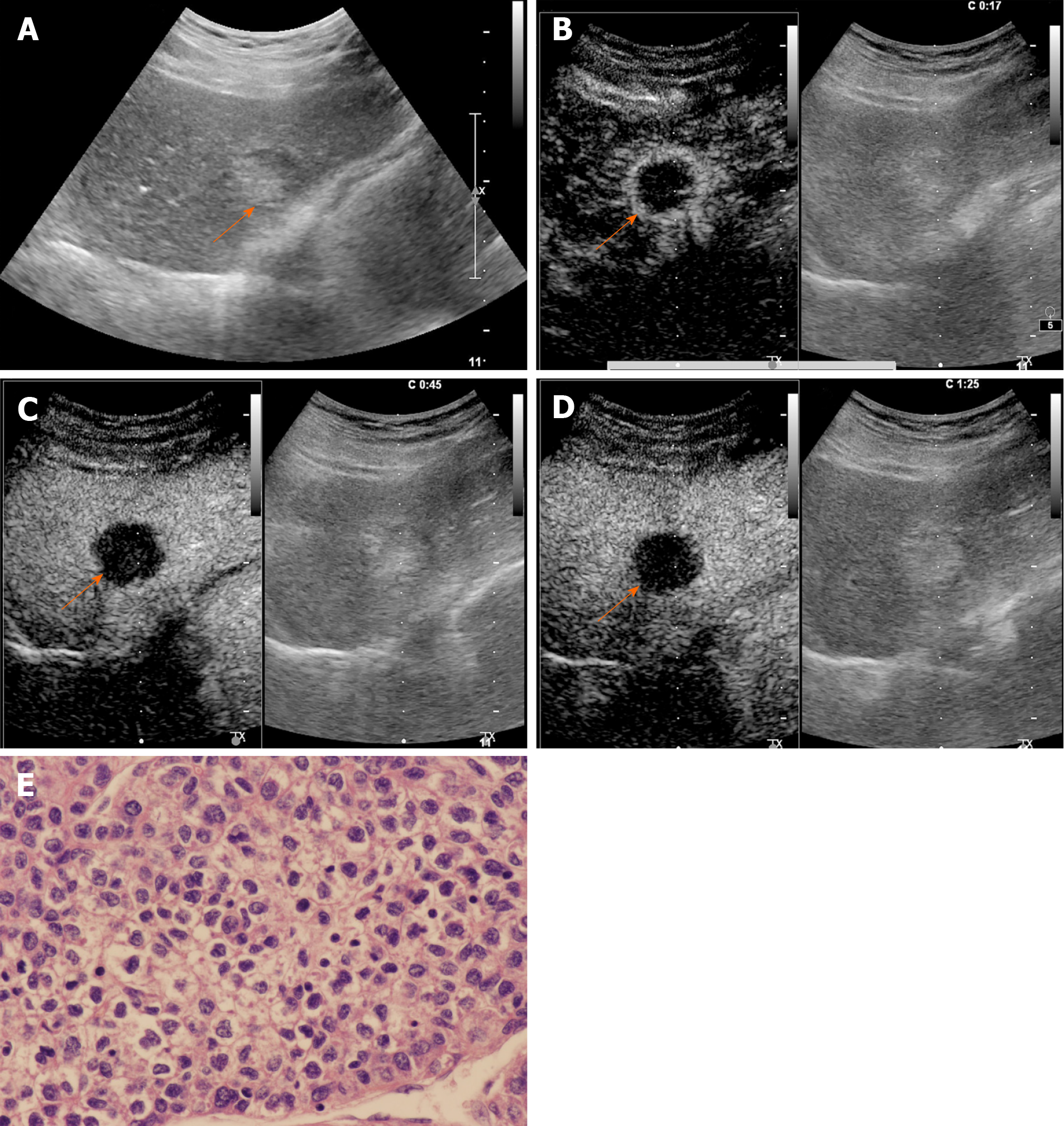
Figure 4 Contrast-enhanced ultrasound examination of a 68-year-old male patient with chronic hepatitis B infection.
A: Conventional grayscale ultrasound demonstrated a mixed echo nodule (arrow) measuring 3.0 cm in diameter in the left lobe of the liver; B: Contrast-enhanced ultrasound illustrated rim arterial phase hyperenhancement (arrow) in the arterial phase; C: Early washout of the contrast agent within 60 s was observed (arrow); D: Late-phase imaging demonstrated marked contrast washout (arrow) within 120 s. The lesion was classified as LR-M due to the aforementioned features; E: The nodule was revealed to be poorly differentiated hepatocellular carcinoma by histopathology (hematoxylin and eosin staining, × 400).
- Citation: Huang JY, Li JW, Ling WW, Li T, Luo Y, Liu JB, Lu Q. Can contrast enhanced ultrasound differentiate intrahepatic cholangiocarcinoma from hepatocellular carcinoma? World J Gastroenterol 2020; 26(27): 3938-3951
- URL: https://www.wjgnet.com/1007-9327/full/v26/i27/3938.htm
- DOI: https://dx.doi.org/10.3748/wjg.v26.i27.3938


