Copyright
©The Author(s) 2020.
World J Gastroenterol. Jul 21, 2020; 26(27): 3929-3937
Published online Jul 21, 2020. doi: 10.3748/wjg.v26.i27.3929
Published online Jul 21, 2020. doi: 10.3748/wjg.v26.i27.3929
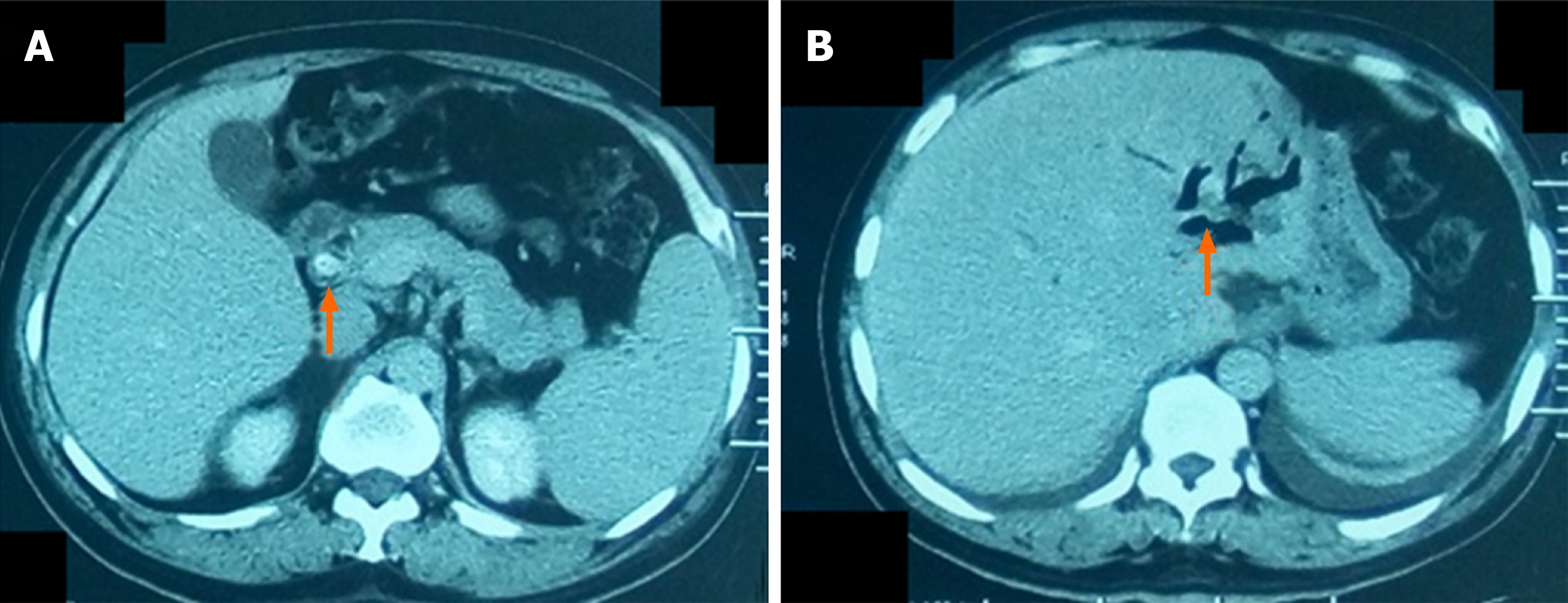
Figure 1 A 58-yr-old man with common bile duct stone and intrahepatic bile duct stone.
A: Computed tomography after contrast administration of the common bile duct stone (arrow); B: Computed tomography without contrast of the intrahepatic bile duct stone (arrow).
- Citation: Liu B, Cao PK, Wang YZ, Wang WJ, Tian SL, Hertzanu Y, Li YL. Modified percutaneous transhepatic papillary balloon dilation for patients with refractory hepatolithiasis. World J Gastroenterol 2020; 26(27): 3929-3937
- URL: https://www.wjgnet.com/1007-9327/full/v26/i27/3929.htm
- DOI: https://dx.doi.org/10.3748/wjg.v26.i27.3929


